


Collection of Urine
Urinalysis is one of the oldest and most informative diagnostic procedures in laboratory medicine. Since urine represents an ultrafiltrate of plasma that has undergone extensive modification through glomerular filtration, tubular reabsorption, secretion, and concentration processes within the nephron, its examination provides valuable information regarding renal function, urinary tract disorders, metabolic diseases, endocrine abnormalities, systemic infections, and toxicological conditions.
The diagnostic value of urinalysis depends not only upon the accuracy of analytical methods but also upon the quality of the specimen submitted for examination. It is estimated that more than 60–70% of laboratory errors occur during the pre-analytical phase, and improper specimen collection is among the most common causes of inaccurate laboratory results. Contamination of urine specimens, incomplete collection, inappropriate storage conditions, bacterial proliferation, degradation of unstable analytes, and failure to use suitable preservatives may significantly alter both qualitative and quantitative test results.
Urine is a biologically unstable fluid. Immediately after voiding, numerous physical, chemical, and microbiological changes begin to occur. Cellular elements such as erythrocytes, leukocytes, and casts undergo progressive degeneration. Bacterial multiplication may result in alkalinization of urine through urea hydrolysis, leading to dissolution of cellular elements and precipitation of crystals. Furthermore, analytes such as glucose, bilirubin, ketone bodies, and urobilinogen may undergo significant degradation during storage. Consequently, strict adherence to standardized collection and preservation protocols is essential to ensure specimen integrity and diagnostic reliability.
The type of urine specimen required depends upon the clinical indication and laboratory investigation requested. Routine screening tests, quantitative biochemical analyses, microbiological cultures, cytological examinations, and specialized endocrinological investigations each require specific collection procedures designed to optimize analytical accuracy.
Collection of Urine for Screening
Routine screening urinalysis constitutes one of the most frequently requested laboratory investigations in clinical practice. It serves as an invaluable tool for the early detection of renal disease, urinary tract infections, diabetes mellitus, hepatobiliary disorders, metabolic abnormalities, and systemic illnesses.
For routine screening, a freshly voided clean urine specimen is generally acceptable. However, whenever possible, an early-morning urine specimen is preferred because it provides the greatest diagnostic yield.
A. Early-Morning Urine Specimen
The first urine voided after an overnight period of sleep is referred to as the early-morning urine specimen. During sleep, fluid intake is absent and renal concentrating mechanisms continue to operate, resulting in the production of a highly concentrated urine specimen. This physiological concentration enhances the detectability of abnormal urinary constituents that may be present only in small quantities during the day.
From a laboratory perspective, the early-morning specimen offers several advantages. The increased concentration of solutes improves the sensitivity of dipstick analysis and microscopic examination. The relatively acidic pH of freshly voided morning urine contributes to the preservation of urinary sediment, thereby facilitating accurate identification of erythrocytes, leukocytes, epithelial cells, casts, and crystals.
Diagnostic Advantages of Early-Morning Urine
- Enhanced Detection of Proteinuria : Protein excretion may fluctuate throughout the day due to posture, physical activity, hydration status, and dietary factors. The concentrated nature of early-morning urine improves the detection of low-grade proteinuria and microalbuminuria, which are important markers of early renal disease.
- Improved Microscopic Examination : The overnight concentration of urine promotes the accumulation of cellular elements and casts, thereby increasing the likelihood of detecting pathological sediment. Hyaline, granular, red cell, and white cell casts are often more readily identified in early-morning specimens than in randomly collected samples.
- Pregnancy Testing : Human Chorionic Gonadotropin (hCG) is generally present in its highest concentration in the first morning specimen. Consequently, early-morning urine is recommended for pregnancy testing, particularly during the early stages of gestation when hormone levels may be relatively low.
- Evaluation of Diabetes Mellitus : Assessment of fasting glycosuria and ketonuria is best performed using the first morning specimen. Detection of glucose and ketone bodies in fasting urine may provide valuable information regarding glycemic control and metabolic status.
- Detection of Orthostatic Proteinuria : Comparison of first-morning urine with daytime specimens may assist in the diagnosis of orthostatic (postural) proteinuria, a benign condition characterized by increased protein excretion during upright posture.
B. Postprandial Urine Specimen
A postprandial urine specimen is collected approximately two to three hours after the consumption of a principal meal. The physiological changes associated with digestion and nutrient absorption may influence the urinary excretion of glucose, amino acids, ketone bodies, and other metabolites.
Historically, postprandial urine glucose testing played an important role in screening for diabetes mellitus before the widespread availability of blood glucose monitoring. Although modern diagnostic criteria are primarily based on plasma glucose and glycated hemoglobin measurements, postprandial urine examination remains useful in selected clinical situations.
Clinical Applications
- Detection of Glycosuria : In individuals with impaired glucose tolerance or diabetes mellitus, blood glucose concentrations may exceed the renal threshold for glucose reabsorption following a meal. Under such circumstances, glucose appears in the urine, producing postprandial glycosuria.
- Evaluation of Carbohydrate Metabolism : Postprandial specimens may reveal transient metabolic abnormalities that are not evident in fasting urine. Therefore, they may provide supplementary information regarding carbohydrate utilization and endocrine function.
- Urobilinogen Estimation : Urobilinogen excretion may exhibit diurnal variation. In certain hepatobiliary disorders, postprandial specimens may improve detection of abnormal urobilinogen concentrations.
- Assessment of Renal Function : Measurement of urinary creatinine and other metabolic products in relation to dietary intake may occasionally be useful in specialized clinical investigations.
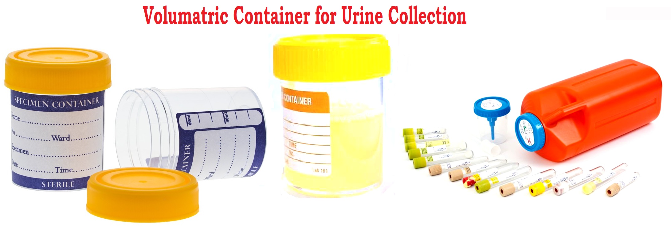
Specimen Containers for Uranalysis
The quality of a urine specimen is influenced not only by the method of collection but also by the characteristics of the collection container. Containers used for routine urinalysis should be:
- Clean, dry, and leak-proof.
- Chemically inert and non-reactive.
- Wide-mouthed to facilitate specimen collection.
- Properly labeled with patient identification details.
- Free from detergents, disinfectants, or chemical residues.
For routine urinalysis, a container with a capacity of approximately 250–300 mL is generally sufficient. The use of sterile containers is recommended whenever microbiological examination is anticipated. Failure to utilize appropriate collection containers may result in contamination, chemical interference, and inaccurate analytical results.
Collection of Urine for Quantitative Analysis
Quantitative urine analysis occupies a central role in clinical laboratory medicine because many biochemical constituents are excreted in urine at varying rates throughout the day. The concentration of an analyte in a single random urine specimen is influenced by hydration status, dietary intake, physical activity, circadian rhythms, medication use, and renal concentrating ability. Consequently, measurement of urinary analytes in a spot specimen may not accurately reflect the total amount excreted by the body.
To overcome these limitations, a complete 24-hour urine collection is often required. This method provides an estimate of the total daily excretion of specific substances and remains the reference procedure for numerous biochemical and endocrinological investigations.
Clinical Significance of 24-Hour Urine
The 24-hour urine specimen provides a comprehensive assessment of renal, metabolic, endocrine, and electrolyte balance by measuring the total quantity of substances excreted over an entire day. The clinical significance of individual parameters is described below. A 24-hour urine analysis provides invaluable information regarding renal function, electrolyte homeostasis, endocrine activity, metabolic status, and stone-forming tendencies. Accurate collection and proper preservation of the specimen are essential for obtaining reliable results and ensuring appropriate clinical interpretation.
A. Renal Function Assessment
- Creatinine Excretion : Creatinine is produced from muscle metabolism at a relatively constant rate and is excreted almost entirely by the kidneys. Measurement of 24-hour urinary creatinine helps assess the completeness of urine collection and provides an estimate of muscle mass. Abnormally low excretion may indicate impaired renal function, reduced muscle mass, malnutrition, or incomplete urine collection, whereas increased excretion may occur in individuals with greater muscle mass or excessive meat intake.
- Creatinine Clearance : Creatinine clearance is a widely used indicator of glomerular filtration rate (GFR). It reflects the kidney’s ability to filter blood and remove waste products. Reduced creatinine clearance is observed in acute and chronic kidney diseases, diabetic nephropathy, glomerulonephritis, and renal vascular disorders. It is useful for staging chronic kidney disease and monitoring disease progression.
- Protein Excretion : Normally, only small amounts of protein are excreted in urine. Increased urinary protein excretion (proteinuria) is a hallmark of glomerular damage and is commonly seen in nephrotic syndrome, diabetic nephropathy, hypertension, lupus nephritis, and chronic kidney disease. Persistent proteinuria is an important predictor of renal disease progression and cardiovascular risk.
- Microalbumin Excretion : Microalbuminuria refers to the excretion of small amounts of albumin that are not detectable by routine urine protein tests. It is an early marker of diabetic nephropathy and hypertensive renal damage. Detection of microalbuminuria allows early intervention to prevent progression to overt kidney disease and is also associated with increased cardiovascular morbidity.
- Urea Excretion : Urea is the major end product of protein metabolism. Measurement of urinary urea excretion helps assess protein intake, nitrogen balance, and renal function. Reduced excretion may occur in renal insufficiency, severe liver disease, or dehydration, while increased excretion may be seen in high-protein diets, catabolic states, fever, or corticosteroid therapy.
B. Electrolyte Studies
- Sodium : Urinary sodium excretion reflects dietary sodium intake, renal tubular function, and fluid balance. It is useful in evaluating dehydration, edema, heart failure, liver cirrhosis, nephrotic syndrome, and disorders of adrenal function. Low urinary sodium may indicate sodium retention, whereas high excretion may occur with diuretic therapy, renal salt-wasting syndromes, and adrenal insufficiency.
- Potassium : Urinary potassium measurement assists in the investigation of hypokalemia and hyperkalemia. Increased urinary potassium excretion may occur in hyperaldosteronism, renal tubular disorders, and diuretic use. Reduced excretion is observed in potassium depletion, adrenal insufficiency, and severe renal failure.
- Chloride : Urinary chloride estimation helps evaluate acid-base disorders and electrolyte imbalances. Low urinary chloride levels are commonly associated with vomiting, metabolic alkalosis, and chloride depletion, whereas elevated levels may occur in renal tubular acidosis and excessive salt intake.
- Calcium : Measurement of urinary calcium is important in the diagnosis and management of nephrolithiasis, hyperparathyroidism, osteoporosis, vitamin D disorders, and metabolic bone diseases. Hypercalciuria increases the risk of kidney stone formation, while reduced excretion may occur in hypoparathyroidism and vitamin D deficiency.
- Phosphate : Urinary phosphate excretion reflects phosphate metabolism and renal tubular handling of phosphate. It is useful in evaluating hyperparathyroidism, chronic kidney disease, vitamin D disorders, rickets, osteomalacia, and bone mineral metabolism abnormalities.
- Magnesium : Urinary magnesium measurement assists in the diagnosis of magnesium deficiency, renal magnesium wasting, and certain endocrine disorders. Increased excretion may occur in diabetes mellitus, alcoholism, and renal tubular diseases, while decreased excretion is seen in dietary deficiency and impaired gastrointestinal absorption.
C. Endocrinological Investigations
- Cortisol : Twenty-four-hour urinary free cortisol measurement is a valuable screening and diagnostic test for hypercortisolism. Increased excretion is characteristic of Cushing’s syndrome and adrenal hyperfunction. It is also useful for monitoring treatment response in patients with cortisol excess.
- Catecholamines : Urinary catecholamine estimation measures epinephrine, norepinephrine, and dopamine excretion. Elevated levels are observed in pheochromocytoma, paraganglioma, severe stress, and certain neuroendocrine disorders. The test aids in the diagnosis of catecholamine-secreting tumors.
- Metanephrines : Metanephrines are metabolites of catecholamines and are considered highly sensitive markers for pheochromocytoma and paraganglioma. Increased urinary excretion strongly suggests the presence of these tumors and is useful in both diagnosis and follow-up.
- Vanillylmandelic Acid (VMA) : VMA is the principal end product of catecholamine metabolism. Elevated urinary VMA levels are associated with pheochromocytoma, neuroblastoma, ganglioneuroma, and other catecholamine-producing tumors. It is frequently used in pediatric oncology and endocrine investigations.
- 5-Hydroxyindoleacetic Acid (5-HIAA) : 5-HIAA is the major metabolite of serotonin. Increased urinary excretion is characteristic of carcinoid tumors, particularly those originating in the gastrointestinal tract. The test is valuable for diagnosis, disease monitoring, and assessment of treatment efficacy.
- Aldosterone : Urinary aldosterone measurement is useful in evaluating disorders of the renin-angiotensin-aldosterone system. Increased excretion occurs in primary hyperaldosteronism (Conn syndrome), secondary hyperaldosteronism, and some forms of hypertension. Reduced levels may be observed in adrenal insufficiency.
D. Metabolic Studies
- Uric Acid Estimation : Urinary uric acid measurement is important in the evaluation of gout, hyperuricemia, kidney stone formation, and disorders of purine metabolism. Increased excretion may occur in leukemia, chemotherapy-induced tumor lysis syndrome, and excessive purine intake.
- Oxalate Determination : Urinary oxalate estimation is essential in investigating recurrent calcium oxalate kidney stones. Elevated excretion may result from primary hyperoxaluria, intestinal malabsorption syndromes, inflammatory bowel disease, or excessive dietary oxalate intake.
- Cystine Excretion : Measurement of urinary cystine is primarily used to diagnose cystinuria, an inherited amino acid transport disorder. Excessive cystine excretion predisposes patients to recurrent cystine stone formation and chronic urinary tract complications.
- Xylose Absorption Test : The urinary excretion of D-xylose following oral administration assesses intestinal absorptive capacity. Reduced urinary excretion indicates malabsorption disorders such as celiac disease, tropical sprue, and other small intestinal mucosal diseases.
- Porphyrin Analysis : Urinary porphyrin estimation plays a crucial role in the diagnosis of porphyrias, a group of inherited or acquired disorders of heme synthesis. Increased porphyrin excretion is associated with acute hepatic porphyrias, cutaneous porphyrias, liver disease, and certain toxic exposures.
Principle of 24-Hour Urine Collection
The objective of a 24-hour collection is to obtain all urine excreted during a complete circadian cycle. Because many biological substances exhibit diurnal variation, collection over a full 24-hour period minimizes fluctuations and provides a more representative measure of daily excretion. The reliability of quantitative urinary measurements depends entirely upon the completeness of specimen collection. Missing even a single voided specimen may lead to significant underestimation of analyte excretion and potentially erroneous clinical interpretation.
Procedure for 24-Hour Urine Collection
The patient should receive detailed written and verbal instructions before beginning the collection.
- Step 1: Initiation of Collection : On the morning of the first day, the patient completely empties the bladder and discards this specimen. The time of voiding is recorded as the official start of the collection period.
- Step 2: Collection Period : All urine subsequently passed during the next 24 hours is collected in the designated container. Special care should be taken to ensure that:
- No specimen is discarded.
- No urine is contaminated with feces, toilet paper, or menstrual blood.
- The collection container remains properly covered.
- Step 3: Completion of Collection : Exactly 24 hours after initiation, the patient empties the bladder one final time and adds this specimen to the collection container. The collection is then complete.
Common Causes of Error in 24-Hour Urine Collection
Errors in quantitative urinalysis are predominantly pre-analytical in nature. Common sources of error include:
- Incomplete Collection : Failure to collect all urine specimens during the collection period remains the most frequent cause of inaccurate results.
- Overcollection : Extending the collection period beyond 24 hours may falsely elevate measured analytes.
- Improper Storage : Bacterial growth and analyte degradation may occur if the specimen is stored at room temperature.
- Incorrect Preservative Use : The use of inappropriate preservatives may interfere with laboratory measurements.
- Recording Errors : Failure to document collection start and end times may invalidate the specimen.
From a laboratory quality management perspective, the completeness of a 24-hour collection may be assessed by measuring total urinary creatinine excretion. Unexpectedly low creatinine values often indicate incomplete collection. Laboratories should reject improperly collected specimens and request recollection whenever specimen integrity is compromised.
Collection of Urine for Bacteriological Examination
Urinary tract infections (UTIs) are among the most common bacterial infections encountered in clinical practice. Accurate microbiological diagnosis requires proper specimen collection because contamination during collection may result in false-positive cultures and inappropriate antimicrobial therapy. The objective of urine culture is to determine whether microorganisms are present within the urinary tract rather than contaminants derived from the external genitalia or distal urethra.
Microbiological Considerations
The distal urethra normally contains commensal microorganisms. During voiding, these organisms may contaminate the urine specimen. Consequently, collection techniques must be designed to minimize contamination while preserving the viability of pathogenic organisms. The preferred specimen for routine bacteriological examination is the clean-catch mid-stream urine (MSU) specimen.
Clean-Catch Mid-Stream Urine
The rationale for collecting the mid-stream portion of urine is based upon the flushing action of the initial urinary stream. The first portion of urine removes:
- Desquamated epithelial cells
- Urethral secretions
- Commensal microorganisms
- External contaminants
The subsequent mid-stream portion more accurately reflects the microbial population present within the bladder.
Patient Preparation
Proper patient education significantly reduces contamination rates and improves diagnostic accuracy.
- Male Patients : The foreskin should be retracted whenever possible, and the glans penis thoroughly cleansed with sterile water or normal saline.
- Female Patients : The labia should be separated during cleansing and maintained apart throughout collection. Cleansing should proceed from anterior to posterior to prevent fecal contamination.
Collection Procedure
- Wash hands thoroughly.
- Cleanse the external genitalia.
- Begin voiding urine.
- Discard the first portion.
- Collect approximately 30–100 mL of the mid-stream urine in a sterile container.
- Avoid touching the inside surface of the container or lid.
- Securely close the container immediately after collection.
Specialized Urine Collection Methods
1. Catheterized Urine Specimen
Catheterization may be required in:
- Critically ill patients
- Patients with urinary retention
- Patients unable to provide a clean-catch specimen
Because catheterization may introduce infection, it should be performed only when clinically indicated.
2. Suprapubic Aspiration
Suprapubic aspiration involves direct needle aspiration of urine from the bladder. This method:
- Produces the least contaminated specimen.
- Is particularly useful in infants and young children.
- Is considered the gold standard for obtaining sterile bladder urine.
Transport and Storage of Urine for Culture
Urine is an excellent growth medium for bacteria. At room temperature:
- Bacterial multiplication may occur rapidly.
- Colony counts may increase artificially.
- Urinary pH may change.
- Cellular elements may deteriorate.
Therefore, specimens should ideally reach the microbiology laboratory within one hour of collection.
If delay is unavoidable:
- Refrigerate at 2–8°C.
- Process within 24 hours whenever possible.
Collection of Mid-Stream Urine
The mid-stream urine (MSU) specimen, also known as a clean-catch urine specimen, is one of the most commonly collected urine samples in clinical laboratories. It is specifically designed to minimize contamination from microorganisms, epithelial cells, mucus, and secretions present in the distal urethra, external genitalia, and surrounding skin. Because contamination can significantly affect the accuracy of urine culture and microscopic examination, proper collection of a mid-stream urine specimen is of utmost importance.
A correctly collected mid-stream urine sample provides a representative specimen of urine from the urinary bladder and is considered the specimen of choice for routine urinalysis, urine culture and sensitivity testing, and the diagnosis of urinary tract infections (UTIs).
During micturition, the initial portion of urine flushes away contaminants present in the distal urethra and external genital region. By discarding the first portion and collecting the middle portion of the urinary stream, the risk of contamination is significantly reduced. The final portion of urine is also discarded to avoid contamination from urethral secretions and residual debris.
Indications for Mid-Stream Urine Collection
Mid-stream urine collection is recommended for:
- Routine urinalysis
- Urine culture and antimicrobial sensitivity testing
- Diagnosis of urinary tract infections
- Investigation of hematuria
- Assessment of proteinuria
- Monitoring of renal diseases
- Screening for asymptomatic bacteriuria
- Follow-up of treated urinary infections
Materials Required
- Sterile wide-mouthed urine container with screw cap
- Soap and clean water or antiseptic cleansing wipes
- Disposable gloves (if required)
- Laboratory request form and patient identification label
Procedure for Collection in Adult Males
- Step 1: Hand Hygiene : The patient should thoroughly wash and dry the hands before specimen collection.
- Step 2: Cleansing of Genital Area : The foreskin should be retracted completely in uncircumcised males. The glans penis and urethral opening should be cleaned with soap and water or sterile cleansing wipes and then allowed to dry.
- Step 3: Initiation of Urination : The patient should begin urinating into the toilet and allow the first portion of urine to pass without collecting it.
- Step 4: Collection of Mid-Stream Sample : Without interrupting the flow of urine, approximately 20–50 mL of the middle portion of the urinary stream should be collected in the sterile container.
- Step 5: Completion :The remaining urine should be voided into the toilet. The container should be immediately capped securely.
- Step 6: Labeling and Transportation : The specimen should be properly labeled with patient identification, date, and time of collection and transported promptly to the laboratory.
Procedure for Collection in Adult Females
- Step 1: Hand Hygiene : Hands should be washed thoroughly before specimen collection.
- Step 2: Separation of Labia : The patient should separate the labia with one hand and keep them separated throughout the collection procedure.
- Step 3: Cleansing : The urethral area should be cleaned from front to back using sterile cleansing wipes or soap and water. This process helps reduce contamination from vaginal and perineal microorganisms.
- Step 4: Begin Urination : The first portion of urine should be voided into the toilet.
- Step 5: Collection of Mid-Stream Portion : Without stopping the urinary stream, the middle portion of urine should be collected directly into the sterile container.
- Step 6: Completion : The remaining urine should be discarded into the toilet, and the container should be capped immediately.
- Step 7: Labeling and Transportation : The specimen should be labeled appropriately and delivered to the laboratory as soon as possible.
Collection in Children
In cooperative children, the clean-catch mid-stream technique should be followed similarly to adults. In infants and young children who are unable to cooperate, alternative collection methods such as catheterization or suprapubic aspiration may be required when sterile specimens are essential for microbiological examination.
Precautions During Collection
- Use only sterile collection containers.
- Avoid touching the inside of the container or lid.
- Do not collect the initial stream of urine.
- Ensure proper cleansing of the genital area before collection.
- Avoid contamination with feces, vaginal secretions, menstrual blood, or toilet paper.
- Collect an adequate volume of urine.
- Close the container immediately after collection.
- Transport the specimen to the laboratory within 1–2 hours.
Advantages of Mid-Stream Urine Collection
- Simple and non-invasive procedure.
- Minimizes contamination from external genital flora.
- Suitable for routine urinalysis and urine culture.
- Provides reliable microbiological results.
- Convenient for outpatient and community-based testing.
The clean-catch mid-stream urine specimen is considered the standard specimen for diagnosing urinary tract infections and evaluating urinary abnormalities. Proper collection significantly reduces contamination, thereby improving the accuracy of microbiological cultures, microscopic examination, and biochemical analysis. A well-collected mid-stream specimen enables clinicians to accurately identify pathogens, assess renal and urinary tract disorders, monitor treatment response, and guide appropriate therapeutic interventions.
The diagnostic value of a urine examination depends greatly on proper specimen collection. A carefully collected mid-stream urine specimen provides the most accurate representation of bladder urine while minimizing contamination from external sources.
Bibliography
- Henry's Clinical Diagnosis and Management by Laboratory Methods. McPherson RA, Pincus MR (Editors). Elsevier; 2021.
- Tietz Fundamentals of Clinical Chemistry and Molecular Diagnostics. Burtis CA, Bruns DE (Editors). Elsevier; 2023.
- Tietz Textbook of Laboratory Medicine. Rifai N, Horvath AR, Wittwer CT (Editors). Elsevier; 2023.
- Clinical Diagnosis and Management by Laboratory Methods. McPherson RA, Pincus MR. Elsevier.
- Graff's Textbook of Urinalysis and Body Fluids. Mundt LA, Shanahan K. Wolters Kluwer/Lippincott Williams & Wilkins; 2015.
- Strasinger's Urinalysis and Body Fluids. Strasinger SK, Di Lorenzo MS. F.A. Davis Company; 2020.
- Clinical Laboratory Medicine. McClatchey KD. Lippincott Williams & Wilkins; 2017.
- Clinical Laboratory Science Review. Jarreau PB. Pearson Education; 2017.
- Clinical and Laboratory Standards Institute. Urinalysis; Approved Guideline. CLSI Document GP16 and current updates.
- International Organization for Standardization. ISO 15189:2022 – Medical Laboratories: Requirements for Quality and Competence.
- Textbook of Medical Laboratory Technology. Praful B. Godkar and Darshan P. Godkar. Bhalani Publishing House; Latest Edition.
- Medical Laboratory Technology Methods and Interpretations. Ramnik Sood. Jaypee Brothers Medical Publishers; Latest Edition.
- World Health Organization. Laboratory Quality Management System (LQMS) Handbook. Geneva: WHO.
- Bishop's Clinical Chemistry. Bishop ML, Fody EP, Schoeff LE. Wolters Kluwer; Latest Edition.
- Practical Clinical Biochemistry. Gowenlock AH, McMurray JR, McLauchlan DM. CBS Publishers & Distributors.