


Synovial Fluid Collection & Analysis : Significance, Collection, Patient preparation, Complication & Laboratory analysis
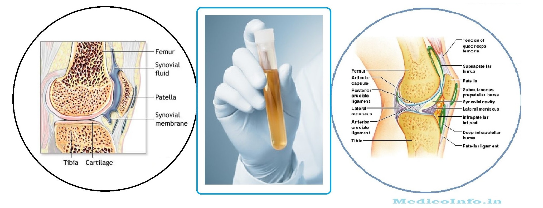
Synovial fluid is the dialysate of plasma with addition of macromolecules; derived from synovium and cartilage. Synovial fluid is found around the joints such as knee, ankle, hip, elbow, wrist and shoulder. The chemical composition of synovial fluid resembles with that of other body fluids such as serous fluids and spinal fluid. In addition, it contains a mucopolysaccharide, hyluronic acid (derived from B cells of synovial membrane), which acts as a binding and protective agent for the connective tissues and also maintaining the high viscosity of the synovial fluid. 
The laboratory examination of this fluid helps to assist in the diagnosis of joint arthritis, gout or infection of the joint (septic arthritis). The synovial fluid cytology is particularly useful noninvasive means of investigating the causes of mono-arthropathies and do distinguish between inflammatory and non-inflammatory joints diseases.
The synovial fluid is obtained by aspiration of a joint. About 1 ml is present in each large joint such as the knee, ankle, hip, elbow, wrist or shoulder. The specimen is obtained only by a trained nurse or by the attending physician. Despatch of the specimen to the laboratory is done as early as possible. Anticogulant is added to the specimen which will be used for cell counting while fluoride is added to the specimen to be used for glucose analysis. The specimen is collected in the following sterile tubes.
- EDTA tube : For cell count and microscopic examination
- Plain tube : For gross examination, mucin clot test, evaluation of viscosity and for microbiological and serological tests.
- Fluoride-oxalate tube : For glucose determination.
Laboratory investigation involves physical examination, microscopic examination, microbiological study and chemical analysis. A few uniques tests include test of viscosity and mucin clot test.
Physical examination
Physical examination ( mainly appearance and clot formation) is done with an Uncentrifuged specimen.
Appearance
The appearance as clear, cloudy, pale yellow, milky, reddish, dark brown, Xanthochromic or greenish. Normal synovial fluid is straw-coloured, clear and viscous, resembling uncooked egg white, and does not clot. The non-inflammatory disorders of the joint usually have clear synovial fluid and one can read newspaper print through the specimen kept in a test tube. Turbidity is usually reported as 1+ to 4+. Turbidity increases in case of inflammatory and infected conditions. All cloudy fluids are however, not inflammatory; further microscopic examination is important, in that crystals, fibrin, amyloid and cartilage fragments, should be sought as these also cause cloudiness. Grossly purulent fluid with an increased leukocyte count is more typical of acute septic arthritis. Presence of large number of crystals or rice bodies may give the fluid a purulent appearance.
Xanthochromia of supernatant synovial fluid indicates an abnormality which may be related to haemophilia, tumours, or trauma. Xanthochromic synovial fluid is difficult to interpret, because of its normal yellow appearance of the synovial fluid; a gross red or dark brown supernatant or the presence of black specks suggests abnormalities. A bloody synovial fluid may be caused by a traumatic tap of the joint and this should not be confused with truly bloody synovial fluid.
[Note: Traumatic tap may cause presence of blood in the synovial fluid. In that case the amount of blood will decrease in the second and the third tube.]
Viscosity test
Synovial fluid is viscous and the viscosity is due to the presence of hyaluronic acid. The viscosity decreases due to the breakdown of hyaluronic acid by the enzyme hyaluronidase in inflammatory disorders.Two tests are performed for detection of viscosity of the synovial fluid - String Test and Mucin clot test.
String Test : Drop the fluid from a syringe and note the length of the tenacious string formed. Use a scale to measure the length. The normal fluid forms a string of at least 4 cm long. If the string breaks before reaching 3 cm length, the viscosity is lower than normal .
Mucin clot test : Hyaluronic acid forms compact clot in the presence of acetic acid. Low concentration of hyaluronic acid does not allow the formation of firm clot.
- Take 20ml of 5%(v/v) acetic acid in a beaker.
- Add 1.0 ml of synovial fluid.
- Observe the formation of clot as follows (a) Firm clot (b) Soft clot (c) Friable clot (d) No clot formation.
- By agitating the solution observe the following.
(a) Clot does not break (b)Clot breaks into small shreds ( since it is poorly formed).
Microscopic Examination
Total leukocyte count and Differential leukocyte count and Wet mount preparation of synovial fluid are very important for the diagnosis of joint related disorders. An anticoagulated (EDTA) specimen is used for the cell count.
Total leukocyte count :
The leukocyte count of normal synovial fluid is about 50 cells/cumm. Normally clear specimen should be used undiluted. Dilute the specimen only when the specimen is turbid. Saline containing methylene blue as diluent for Total leukocyte count. Don't use diluted Acetic acid as the diluent for WBC count. Normal WBC diluting fluid contain acetic acid precipitate the hyaluronic acid-protein complex.
[Note : In the case of a bloody specimen, use o.1N HCL in saline or 1% Saponin in saline as a diluent. In the case of inflammatory disorders the white cell count is high (>10,000/cu mm).]
Differential WBC Count :
Prepare a thin smear of the sediment and staining the smear with Wright-Giemsa stain . An anticoagulated (EDTA) specimen is used for the Differential leukocyte count. Normal fluid contains about 25% of polymophonuclear cells. Increase in these cells (above 70%) indicate bacterial arthritis. Moderate increase in neutrophils ( 40 to 60%) is observed in rheumatic fever, gout, tuberculous arthritis and in rheumatic arthritis.
Wet mount preparation :
Prepare a wet coverslip preparation of the synovial fluid with alcohol and acetone. Observe the slide first under low power objective and afterwards under high power objective. The various observations are as follows :
| Observation | Condition |
| Urate crystals (Needle shaped, Intracellular) | Gouty arthritis |
| Rhomboid calcium pyrophosphate crystals | Pseudogout |
| Cholesterol crystals (Flat, clear, rhombic appearance with one corner punched out | Rheumatoid arthritis |
[Note : Perform Gram's staining and acid fast staining on the sediment smears.]
Chemical Examination
Chemical examination of Synovial fluid (particularly glucose) play an important role for detect the nature of joint related diseases. In case of non-inflammatory arthritis, the difference of blood glucose and synovial fluid glucose is only 10 mg/dl which increases to 25 - 50 mg/dl in case of infectious septic arthritis. Under mild inflammatory condition as like Rheumatoid arthritis, gout or Pseudogout the glucose contain of the synovial fluid is close to normal but increases with severe inflammation. The latter is also associated with increases of protein contain). Benedict quantitative test, Orthotoluidine test and GOD-POD enzymatic methods are used for determination of glucose concentration of Synovial fluid.