


Atrial Fibrillation (AFib) – Sign and Symptoms, Risk Factors, Diagnosis, Complications, Treatment and Prevention
Atrial fibrillation (AFib or AF) is the most common sustained cardiac arrhythmia encountered in clinical practice, affecting an estimated 37–44 million individuals worldwide and representing a major global public health burden. It is characterized by chaotic, disorganized electrical activity within the atria, replacing the normal coordinated depolarization generated by the sinoatrial (SA) node with rapid, irregular, and unsynchronized electrical impulses firing at rates of 350–600 impulses per minute. The resulting ventricular response is irregularly irregular, producing the hallmark clinical finding of an irregularly irregular pulse, variable pulse amplitude, and frequently symptomatic palpitations, dyspnea, and exercise intolerance. 
The pathophysiology of AFib involves a complex interplay of electrical and structural remodeling of the atrial myocardium. Triggers — most commonly arising from pulmonary vein ostia — initiate episodes of AF, which are then sustained by a combination of re-entrant wavelets (multiple-wavelet hypothesis), rotors (spiral waves), focal ectopic discharge, and calcium dysregulation within atrial cardiomyocytes. Structural remodeling, including atrial fibrosis and dilation, creates a favorable substrate for re-entry. Inflammatory cytokines, oxidative stress, renin-angiotensin-aldosterone system activation, and autonomic dysregulation further contribute to atrial remodeling and arrhythmia perpetuation. The principle of "AFib begets AFib" — where AF-induced electrical and structural remodeling promotes the transition from paroxysmal to persistent to permanent forms — underpins the progressive nature of the disease.
Atrial Fibrillation is classified by temporal pattern into first detected, paroxysmal (<7 days, self-terminating), persistent (>7 days), long-standing persistent (>12 months), and permanent AF. Risk factors span cardiovascular (hypertension, coronary artery disease, heart failure, valvular heart disease), systemic (diabetes mellitus, obesity, obstructive sleep apnea, chronic kidney disease, hyperthyroidism), and lifestyle factors (alcohol, physical inactivity, smoking). The most feared complication of AF is thromboembolic stroke, resulting from stasis-induced thrombus formation, predominantly in the left atrial appendage (LAA), with an approximately 5-fold increased stroke risk compared to the general population. Other complications include heart failure, tachycardia-induced cardiomyopathy, cognitive decline, dementia, and significantly impaired quality of life.
Management of Atrial Fibrillation is built upon three interlocking pillars: anticoagulation for stroke prevention (guided by CHA₂DS₂-VASc scoring), rate control (target resting ventricular rate <110 bpm), and rhythm control (restoration and maintenance of sinus rhythm via pharmacological cardioversion, antiarrhythmic drugs, electrical cardioversion, or catheter ablation). The landmark EAST-AFNET 4 trial demonstrated superior cardiovascular outcomes with early rhythm control initiated within 12 months of AF diagnosis, shifting paradigms toward earlier intervention. Direct oral anticoagulants (DOACs — apixaban, rivaroxaban, dabigatran, edoxaban) have largely replaced warfarin as the preferred stroke prophylaxis agents due to superior efficacy-safety profiles. Catheter ablation (primarily pulmonary vein isolation) offers rhythm control with superior freedom from AF recurrence compared to antiarrhythmic drug therapy in appropriate candidates. Prevention strategies targeting modifiable risk factors — particularly hypertension, obesity, sleep apnea, and alcohol reduction — are increasingly recognized as essential components of a comprehensive "upstream therapy" approach to AFib management.
Classification of Atrial Fibrillation
AFib is classified by temporal pattern (reflecting disease progression), clinical setting, and underlying etiology. The ESC 2020 and AHA/ACC 2023 guidelines use the following classification framework:
| Classification | Definition | Clinical Implication |
|---|---|---|
| First Detected AF | First diagnosed episode, regardless of duration or symptoms | Underlying cause must be identified; may be paroxysmal or persistent |
| Paroxysmal AF | Self-terminating within 7 days (usually <48 hours) | Recurrent; sinus rhythm restoration is spontaneous; ablation curative in many |
| Persistent AF | Sustained >7 days or requiring cardioversion to terminate | Requires active rhythm or rate control strategy; atrial remodeling underway |
| Long-standing Persistent AF | Continuous AF >12 months when rhythm control is pursued | Significant structural remodeling; ablation less successful |
| Permanent AF | AF accepted by patient and clinician; rhythm control no longer pursued | Rate control + anticoagulation strategy; decision can be revisited |
| Valvular AF | AF with moderate-to-severe mitral stenosis or mechanical prosthetic valve | Warfarin (not DOACs) mandatory for anticoagulation |
| Non-valvular AF | AF in absence of rheumatic mitral stenosis or prosthetic heart valves | DOACs preferred for stroke prophylaxis |
| Lone AF | AF in patients <60 years with no structural heart disease or hypertension | Low thromboembolic risk; clinical significance disputed in modern guidelines |
| Subclinical / Silent AF | AF detected incidentally by implanted devices or monitoring; no symptoms | Carries stroke risk; anticoagulation decision based on CHA₂DS₂-VASc score |
The 4S-AF scheme (ESC 2020) provides a structured assessment framework: Stroke risk, Symptom severity (EHRA score I–IVb), Severity of AF burden (temporal classification), and Substrate severity (structural heart disease, comorbidities) — guiding integrated, patient-centered management decisions.
Signs and Symptoms of Atrial Fibrillation
The clinical presentation of AFib is highly variable, ranging from completely asymptomatic (silent AF) to severely symptomatic with hemodynamic compromise. Symptom severity depends on the ventricular rate response, underlying cardiac function, duration of AF, and individual patient physiology. The EHRA (European Heart Rhythm Association) symptom classification standardizes the assessment of AF-related symptom burden. 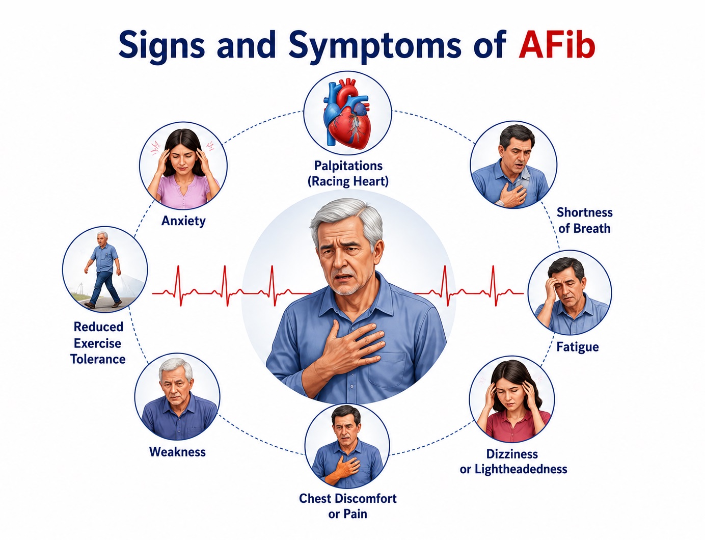
1. Cardiac Symptoms
- Palpitations: The most commonly reported symptom, experienced as an uncomfortable awareness of an irregular, fast, or forceful heartbeat. Described variably as "fluttering," "racing," "skipping," or "flopping" sensations in the chest. Palpitations reflect the irregularly irregular ventricular rate response to disordered atrial electrical activity and often prompt the initial medical evaluation. They may be continuous or episodic, corresponding to the paroxysmal or persistent nature of AF.
- Dyspnea and exertional breathlessness: Commonly reported as the dominant symptom, particularly in patients with underlying structural heart disease or compromised ventricular function. Dyspnea arises from impaired cardiac output due to loss of atrial systolic contribution to ventricular filling ("atrial kick"), tachycardia-induced diastolic dysfunction, and elevated left atrial pressure with secondary pulmonary venous congestion. In patients with preserved ejection fraction, rapid ventricular rates during AF particularly impair diastolic filling time.
- Chest pain or pressure: Chest discomfort in AF may reflect tachycardia-induced subendocardial ischemia in patients with coronary artery disease, increased myocardial oxygen demand from the elevated heart rate, or anxiety. Typical ischemic chest pain with AF warrants urgent exclusion of concurrent acute coronary syndrome.
- Reduced exercise tolerance: Even mildly symptomatic patients experience measurable impairment in functional capacity. The loss of atrial systolic function reduces cardiac output by approximately 15–30%, particularly during exercise when atrial contribution to ventricular filling becomes increasingly important. Patients report inability to perform activities previously well-tolerated.
- Fatigue and weakness: Chronic fatigue is among the most pervasive and functionally limiting AF symptoms, attributed to reduced cardiac output, impaired chronotropic response to exercise, sleep disruption from nocturnal palpitations, and the psychological burden of living with a chronic arrhythmia. Fatigue often persists even after restoration of sinus rhythm, reflecting underlying comorbidities and deconditioning.
2. Neurological and Cerebrovascular Symptoms
- Stroke and transient ischemic attack (TIA): AF-associated thromboembolism from left atrial appendage (LAA) thrombi is responsible for approximately 20–30% of all ischemic strokes. AF-related strokes tend to be cardioembolic, with large vessel occlusion causing severe neurological deficits. Symptoms include sudden onset facial asymmetry, unilateral arm or leg weakness, speech disturbance (aphasia or dysarthria), sudden vision loss, and severe headache. Patients with previously undiagnosed ("silent") AF may first present with an embolic stroke.
- Cognitive impairment and dementia: Growing evidence demonstrates a strong, independent association between AFib and cognitive decline, vascular dementia, and Alzheimer-type dementia, even in the absence of clinically overt stroke. Mechanisms include silent cerebral microemboli, cerebral hypoperfusion from irregular cardiac output, and inflammatory cytokine-mediated neuroinflammation. Patients may report subjective memory complaints, difficulty concentrating, and reduced processing speed.
- Dizziness and presyncope: Lightheadedness, disequilibrium, and near-fainting episodes arise from fluctuating cerebral perfusion due to the irregularly irregular cardiac rhythm and variable beat-to-beat cardiac output. Episodes often correlate with periods of particularly rapid or slow ventricular response.
- Syncope: Transient loss of consciousness occurs in approximately 3–5% of AFib patients, resulting from acute hemodynamic compromise — either from very rapid ventricular rates reducing cardiac output, or from a pause following spontaneous termination of AF (post-conversion pause, particularly in sick sinus syndrome). Syncope in AF mandates urgent investigation for underlying accessory pathway conduction (Wolff-Parkinson-White syndrome) where pre-excited AF can degenerate to ventricular fibrillation.
3. Autonomic and Systemic Symptoms
- Anxiety and psychological distress: AFib exerts a profound psychological burden. The unpredictability of paroxysmal episodes, fear of stroke, medication side effects, and the chronic nature of the condition generate significant anxiety, depression, and health-related quality of life impairment. Psychological distress may paradoxically trigger AF episodes through autonomic nervous system activation and adrenergic surges, creating a bidirectional relationship.
- Polyuria: Rapid atrial distension and elevated atrial wall tension during AF stimulate secretion of atrial natriuretic peptide (ANP) and brain natriuretic peptide (BNP) from atrial cardiomyocytes. These natriuretic peptides promote renal sodium and water excretion, resulting in clinically noticeable polyuria during and immediately after AF episodes — a symptom that may help patients recognize the onset and termination of paroxysmal AF.
- Diaphoresis: Profuse sweating during AF episodes reflects heightened sympathetic nervous system activation, elevated catecholamine levels, and the hemodynamic stress imposed by rapid, irregular ventricular rates.
- Gastrointestinal discomfort: Nausea, epigastric discomfort, and reduced appetite may accompany AF episodes, attributed to autonomic dysregulation, reduced splanchnic blood flow, and — in some patients — medication side effects from antiarrhythmic or anticoagulation therapy.
4. Signs on Physical Examination
- Irregularly irregular pulse: The pathognomonic clinical sign of AFib on physical examination — a completely irregular pulse with variable amplitude and rate, in contrast to the regular irregularity of second-degree AV block or other arrhythmias. Appreciated at the radial or carotid pulse, with variable intensity of the apical impulse on cardiac auscultation.
- Pulse deficit: A difference between the apical heart rate (auscultated at the cardiac apex) and the peripheral radial pulse rate, resulting from inadequately filled left ventricular contractions following short R-R intervals that generate insufficient stroke volume to produce a palpable peripheral pulse wave.
- Irregular heartbeat on auscultation: Completely irregular heart sounds without discernible pattern on cardiac auscultation. The absence of a fourth heart sound (S4 — which requires atrial systole) is consistent with AF. A variable intensity first heart sound (S1) reflects changing pre-systolic atrial pressure and variable ventricular preload.
- Signs of heart failure: In decompensated AF or tachycardia-induced cardiomyopathy: elevated jugular venous pressure, bilateral basal crackles (pulmonary edema), peripheral pitting edema, hepatomegaly, and a displaced apex beat reflecting left ventricular enlargement.
- Signs of hyperthyroidism: In AF secondary to thyrotoxicosis: tremor, exophthalmos, goiter, warm moist skin, and hyperreflexia — a potentially reversible etiology requiring thyroid function testing in all new AF presentations.
- Tachycardia: Rapid ventricular rate response (>100 bpm) in uncontrolled AF, or bradycardia if rate-controlling medications are effective or sick sinus syndrome co-exists.
5. Asymptomatic (Silent) AFib
Up to 25–40% of Atrial Fibrillation episodes are entirely asymptomatic, particularly in elderly patients, those with longstanding persistent AF, and individuals with reduced perception of cardiac rhythm changes. Silent AF is often detected incidentally on routine ECG, exercise testing, ambulatory monitoring, or through cardiac implantable electronic devices (CIEDs) that log arrhythmic episodes as "atrial high-rate episodes" (AHREs). The importance of silent AF lies in its equivalent thromboembolic risk — patients with device-detected AF maintain a significantly elevated stroke risk despite absence of symptoms, necessitating anticoagulation decisions based on CHA₂DS₂-VASc scoring regardless of symptomatic status.
Risk Factors of Atrial Fibrillation
Atrial Fibrillation (AFib) is a multifactorial arrhythmia arising from the interaction of multiple modifiable and non-modifiable risk factors that collectively promote atrial electrical instability and structural remodeling. Recognition and targeted modification of risk factors constitute a critical upstream prevention and management strategy. 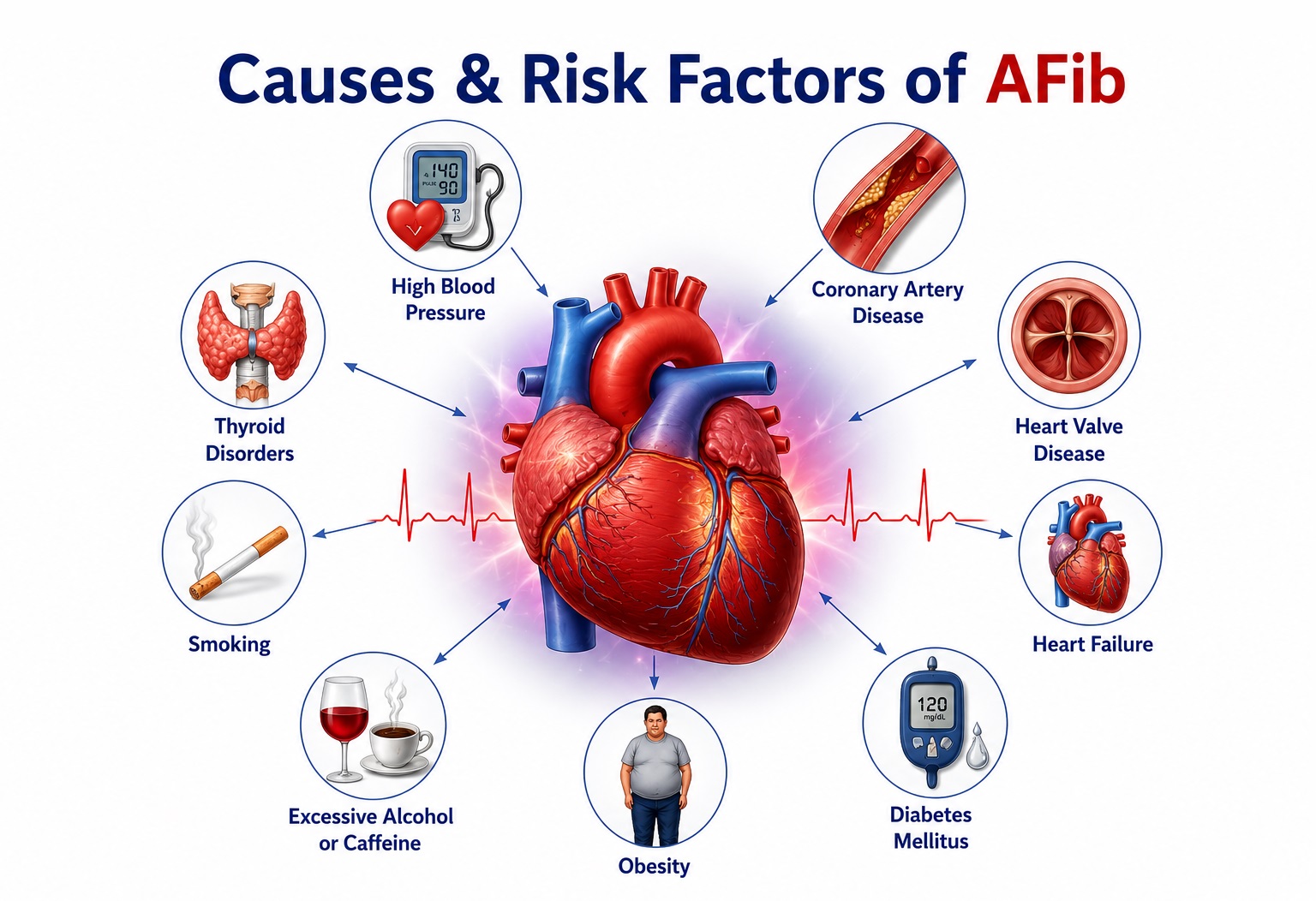
1. Cardiovascular Risk Factors
- Hypertension (systemic arterial hypertension): The single most prevalent and attributable risk factor for Atrial Fibrillation at the population level, responsible for approximately 14–22% of all AFib cases. Sustained elevation of systemic blood pressure imposes chronic pressure overload on the left atrium, promoting atrial dilation, fibrosis, and electrical remodeling that creates a substrate for AF initiation and perpetuation. Even prehypertension confers measurable AF risk. Optimal blood pressure control is associated with a 35–40% reduction in AF risk and is foundational to upstream therapy.
- Heart failure: Bidirectionally related to Atrial Fibrillation — heart failure promotes AF through elevated left atrial filling pressures, atrial stretch, neurohormonal activation (RAAS, sympathetic nervous system), and atrial fibrosis; conversely, AF precipitates and worsens heart failure through tachycardia-induced cardiomyopathy and loss of atrial contribution to cardiac output. AF is present in approximately 30–40% of heart failure patients, rising to >50% in advanced (HFrEF stage D) disease.
- Coronary artery disease (CAD): Myocardial ischemia and infarction promote atrial electrical instability through altered autonomic tone, inflammatory mediator release, and structural changes including atrial dilation from impaired left ventricular function. AF complicates approximately 6–10% of acute myocardial infarctions and is associated with worsened prognosis in this context.
- Valvular heart disease: Mitral valve disease — particularly mitral stenosis and mitral regurgitation — elevates left atrial pressure and volume, driving atrial dilation and fibrosis. Rheumatic mitral stenosis is the classic valvular cause of AF (valvular AF). Tricuspid valve disease similarly causes right atrial enlargement and AF. Aortic stenosis and regurgitation promote AF through secondary left ventricular hypertrophy and diastolic dysfunction.
- Cardiomyopathies: Hypertrophic cardiomyopathy (HCM) carries a particularly elevated AF risk (~20–25% lifetime prevalence) due to diastolic dysfunction and left atrial hypertension. Dilated cardiomyopathy and infiltrative cardiomyopathies (amyloidosis, sarcoidosis) also predispose to AF through atrial remodeling and fibrosis.
- Congenital heart disease: Adults with repaired or unrepaired congenital cardiac lesions — particularly atrial septal defects, Fallot tetralogy, Fontan circulation, and Ebstein anomaly — have significantly elevated AF and atrial flutter risk due to chronic atrial dilation, pressure-volume loading, and surgical scar-related re-entry circuits.
2. Systemic and Metabolic Risk Factors
- Diabetes mellitus: Independent risk factor for AFib, conferring approximately 34–40% increased risk. Mechanisms include autonomic neuropathy altering atrial electrophysiology, oxidative stress promoting atrial fibrosis, inflammatory cytokine release, and endothelial dysfunction. Poorly controlled glycemia exerts more pronounced AF risk than well-controlled diabetes, highlighting the importance of metabolic management.
- Obesity: Strongly associated with AF risk, with a 49% increased risk per 5-unit BMI increment. Adipose tissue — particularly pericardial and epicardial fat — exerts direct paracrine inflammatory effects on adjacent atrial myocardium, promotes adipokine-mediated fibrosis, and increases left atrial volume through mechanical effects of obesity on cardiac structure. The LEGACY trial demonstrated that progressive and sustained weight loss (>10% body weight) significantly reduces AF burden and symptom severity.
- Obstructive sleep apnea (OSA): A highly prevalent but underdiagnosed AF risk factor. OSA promotes AF through repetitive intermittent hypoxia causing autonomic surges, sympathetic activation, hypoxia-induced atrial remodeling, systemic inflammation, and elevated intrathoracic pressure fluctuations that stretch the pulmonary veins and left atrium. CPAP therapy reduces AF recurrence and should be optimized before ablation procedures.
- Hyperthyroidism: Both overt and subclinical hyperthyroidism increase AF risk. Excess thyroid hormone accelerates automaticity in atrial cardiomyocytes, shortens atrial refractory periods, and enhances adrenergic sensitivity. AF affects approximately 10–15% of hyperthyroid patients and 1–2% of subclinical hyperthyroid individuals. Thyroid function testing is mandatory in all new AFib presentations as hyperthyroidism-induced AF may be reversible with treatment of the underlying thyroid disorder.
- Chronic kidney disease (CKD): Independent risk factor mediated through volume overload, uremic toxin-induced atrial remodeling, inflammation, electrolyte dysregulation (hyperkalemia, hypomagnesemia), secondary hypertension, and secondary hyperparathyroidism. AF prevalence increases with CKD stage, reaching over 20% in end-stage renal disease (ESRD). Management is complicated by bleeding risk from uremia and altered anticoagulant pharmacokinetics in renal impairment.
- Hyperlipidemia and metabolic syndrome: Dyslipidemia contributes to AF risk through systemic inflammation, oxidative stress, and direct membrane effects on ion channel function. The metabolic syndrome — clustering hypertension, diabetes, obesity, and dyslipidemia — confers substantially amplified AF risk through multiple mechanistic pathways.
3. Lifestyle and Environmental Risk Factors
- Alcohol consumption: Both heavy chronic alcohol use and acute binge drinking ("holiday heart syndrome") precipitate AF. Alcohol exerts direct toxic effects on atrial myocardium, promotes oxidative stress and inflammation, activates the sympathetic nervous system, and shortens atrial refractory periods. A dose-dependent relationship exists; even moderate habitual alcohol consumption (2–3 standard drinks per day) significantly increases AF risk, and complete alcohol abstinence in patients with AF has been shown to reduce AF recurrence in the REDUAF trial.
- Smoking: Active smoking increases AF risk by approximately 50% compared to non-smokers. Tobacco smoke constituents promote systemic inflammation, endothelial dysfunction, oxidative stress, and autonomic dysregulation, collectively predisposing to atrial remodeling and arrhythmia. Former smokers retain an elevated but declining risk compared to current smokers.
- Excessive physical activity: Paradoxically, while regular moderate exercise is cardioprotective, endurance athletic training — particularly long-term high-intensity exercise such as marathon running, long-distance cycling, and cross-country skiing — is associated with a 5-fold increased AF risk in males. Mechanisms include vagally mediated sinus bradycardia (creating favorable conditions for atrial ectopy), atrial dilation from chronic volume loading, and inflammatory remodeling from repetitive exercise-induced atrial stretch.
- Sedentary lifestyle: Physical inactivity and excessive sedentary time are independently associated with elevated AF risk, mediated through obesity, hypertension, metabolic syndrome, and reduced cardiorespiratory fitness.
- Caffeine: The historical concern that caffeine promotes AF is not supported by current evidence. Moderate coffee and tea consumption does not increase AF risk and may be mildly protective. High-dose supplemental caffeine, however, should be avoided in susceptible individuals.
4. Non-Modifiable and Genetic Risk Factors
- Advanced age: The strongest single risk factor for Atrial Fibrillation. Atrial Fibrillation prevalence rises exponentially with age: from 0.1% in those under 55 years to >9% in those over 80 years. Aging promotes atrial fibrosis, cellular ion channel dysfunction, reduced conduction velocity, and accumulation of structural remodeling — collectively creating an ideal electrophysiological substrate for AF initiation and maintenance.
- Male sex: Males have a 1.5–2.0-fold higher AF incidence than females across all age groups. However, females with AF tend to experience more severe symptoms, higher stroke risk, and worse quality of life, possibly related to hormonal influences on atrial electrophysiology and delayed diagnosis.
- Family history and genetic predisposition: First-degree relatives of AFib patients have a 40–85% increased AF risk. Genome-wide association studies (GWAS) have identified over 100 genetic loci associated with AF risk, including variants in genes encoding cardiac ion channels (SCN5A, KCNQ1, KCNH2), gap junction proteins (connexin 40 — GJA5), and transcription factors (PITX2, ZFHX3, TBX5). Rare Mendelian forms of familial AF are caused by monogenic mutations in cardiac ion channel genes.
- Race and ethnicity: AF is more prevalent and is diagnosed earlier in Caucasians compared to Black and Asian populations, despite Black patients having higher rates of hypertension and other AF risk factors. Black patients with AF experience higher stroke rates and worse outcomes, possibly related to healthcare access disparities and higher rates of uncontrolled hypertension.
5. Acute Precipitants and Reversible Causes
- Post-cardiac surgery AF (POAF): AF occurs in 20–40% of patients following cardiac surgery (CABG, valve surgery) and up to 60% after pericardiotomy procedures. Mechanisms include surgical trauma, pericardial inflammation, oxidative stress, sympathetic activation, and electrolyte shifts. POAF is often self-limiting but increases the risk of stroke and prolonged hospitalization.
- Acute illness: Sepsis, pneumonia, pulmonary embolism, acute myocardial infarction, and pericarditis are common acute precipitants of new-onset AF, mediated through hypoxia, systemic inflammation, catecholamine surge, and autonomic activation.
- Electrolyte abnormalities: Hypokalemia, hypomagnesemia, and hypocalcemia alter atrial action potential characteristics, increasing susceptibility to triggered activity and re-entry. Electrolyte correction is mandatory before cardioversion.
- Drugs and toxins: Sympathomimetic agents (cocaine, amphetamines, ephedrine), theophylline, digoxin toxicity, and certain antiarrhythmic drugs may precipitate or maintain AF.
Diagnosis of Atrial Fibrillation
Diagnosis of Atrial Fibrillation requires integration of clinical assessment, electrocardiographic confirmation, and structured evaluation to determine AF type, hemodynamic impact, underlying etiology, thromboembolic risk, and suitability for rate versus rhythm control strategies.
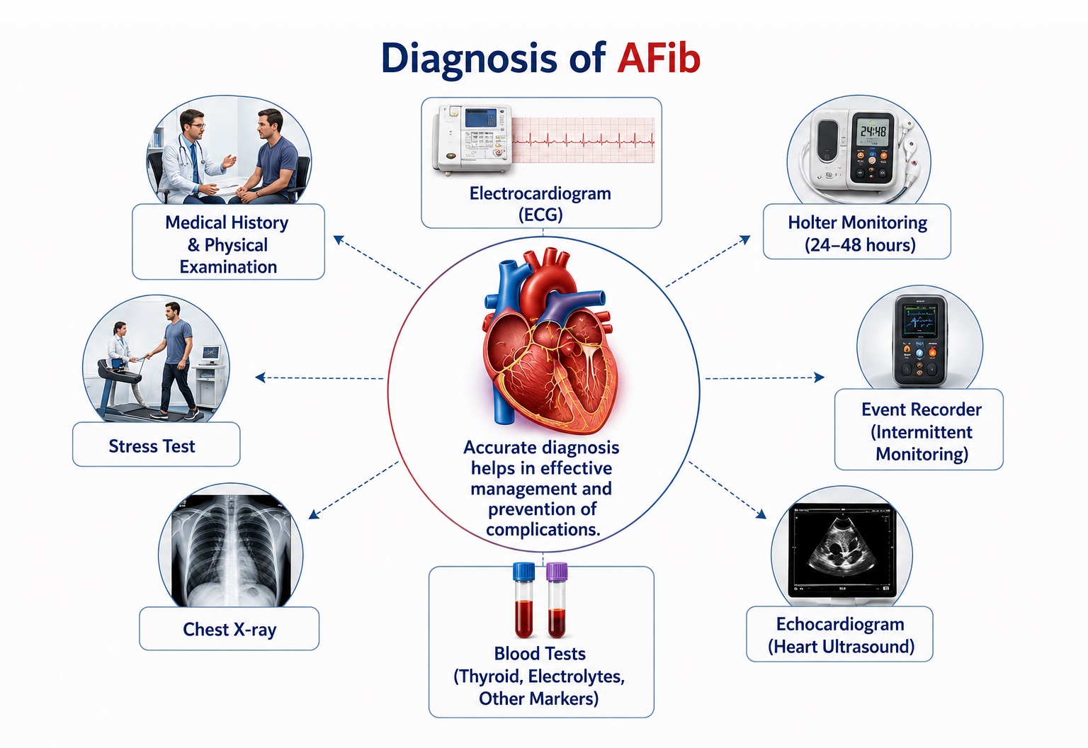
1. Clinical Examination
- Symptom characterization: Onset, duration, frequency, and pattern of palpitations, dyspnea, and other symptoms. Whether symptoms are continuous or episodic, and any identified triggers (exercise, alcohol, emotional stress, caffeine, positional changes).
- EHRA symptom classification: Standardized assessment of symptom severity (EHRA I–IVb) to guide treatment intensity and rhythm control decisions.
- Cardiovascular history: Prior history of hypertension, coronary artery disease, heart failure, valvular disease, prior stroke or TIA, diabetes mellitus, thyroid disease, obesity, and sleep apnea.
- Medication review: Current and prior antiarrhythmic drugs, anticoagulants, rate-controlling agents, and potential AF precipitants (sympathomimetics, theophylline).
- Lifestyle factors: Alcohol intake (quantity, pattern), physical activity level, tobacco use, and recreational drug use.
- Family history: First-degree relatives with AF, hereditary arrhythmia syndromes, sudden cardiac death, or cardiomyopathies.
- Vital signs: Heart rate (ventricular rate response), blood pressure (both arms for asymmetry suggesting subclavian stenosis), respiratory rate, oxygen saturation, and temperature.
- Cardiovascular examination: Irregularly irregular pulse (radial and carotid), pulse deficit assessment, cardiac auscultation (irregular heart sounds, murmurs suggesting valvular disease, S3 gallop in heart failure), jugular venous pressure evaluation, assessment for peripheral edema.
- Thyroid examination: Goiter, exophthalmos, tremor, skin changes — mandatory in new AF presentations.
- Respiratory examination: Crackles suggesting pulmonary congestion or pneumonia-precipitated AF.
- Neurological examination: Focal deficits suggesting concurrent cardioembolic stroke or TIA.
2. Electrocardiography (ECG)
The 12-lead ECG is the gold-standard diagnostic tool for Atrial Fibrillation (AFib). Characteristic features include:
- Absence of distinct P waves: Normal P waves are replaced by rapid, irregular, low-amplitude fibrillatory (f) waves — best visualized in leads V1, II, III, and aVF — with a rate of 350–600 per minute. These f waves vary in amplitude, morphology, and interval, producing the characteristic baseline undulation.
- Irregularly irregular R-R intervals: The most clinically obvious ECG feature — completely irregular ventricular complexes without any discernible pattern, in contrast to the regular irregularity of other arrhythmias such as second-degree AV block.
- Variable R-R intervals: No two consecutive R-R intervals are equal, reflecting the random, variable conduction of AF impulses through the AV node.
- Narrow QRS complexes (usually): QRS complexes are typically narrow (<120 ms) unless aberrant ventricular conduction (LBBB, RBBB), rate-dependent aberrancy, or pre-excitation (Wolff-Parkinson-White syndrome) is present. Wide complex AF mandates urgent exclusion of pre-excited AF.
- Associated findings: Left ventricular hypertrophy (hypertension), left atrial enlargement pattern, ST-segment changes suggesting ischemia, and evidence of pre-excitation (delta waves) may be identified.
- Rapid ventricular rate (>100 bpm): Suggests inadequate rate control or absence of AV nodal blocking therapy; bradycardic response may indicate AV nodal disease or excess rate-controlling medication.
3. Ambulatory ECG Monitoring
- 24–48 hour Holter monitoring: Standard ambulatory ECG recording for detection of paroxysmal AF in patients with intermittent symptoms or to quantify AF burden (% time in AF) in persistent/longstanding AF. Sensitivity is limited by recording duration relative to AF episode frequency.
- 7-day event recorder or extended Holter (14–30 days): Significantly increases AF detection yield for infrequent paroxysmal episodes. Recommended for patients with unexplained stroke and negative initial monitoring, or for symptom-rhythm correlation in symptomatic patients.
- Implantable loop recorder (ILR): Subcutaneously implanted continuous cardiac monitor providing up to 3 years of AF monitoring. Highly effective for cryptogenic stroke evaluation, detecting AF in 12–30% of patients with unexplained stroke over 3 years of monitoring. Also used for monitoring AF burden after ablation.
- Wearable and digital health monitoring: Photoplethysmography (PPG)-based smartwatch algorithms, single-lead ECG patches (KardiaMobile, Zio patch), and AI-powered ECG interpretation tools enable scalable population-level AF screening. Validated algorithms demonstrate 87–98% sensitivity for AF detection in screening contexts.
4. Echocardiography
- Transthoracic echocardiography (TTE): Mandatory in all newly diagnosed Atrial Fibrillation patients to evaluate left ventricular size and function (systolic and diastolic), valvular morphology and function, wall motion abnormalities, right heart structure and pressure, pericardial disease, and atrial dimensions. Findings guide treatment decisions — HF with reduced EF mandates specific medical therapy, valvular AF alters anticoagulation strategy, and structural heart disease modifies ablation suitability.
- Left atrial diameter and volume: Left atrial enlargement (LA diameter >4.0 cm or LA volume index >34 mL/m²) is both a consequence and predictor of AF and identifies patients at higher risk of AF recurrence after cardioversion or ablation.
- Transoesophageal echocardiography (TOE/TEE): Gold standard for exclusion of left atrial appendage (LAA) thrombus prior to cardioversion when adequate anticoagulation cannot be confirmed (AF duration >48 hours or unknown). TOE provides superior visualization of the LAA compared to TTE, with sensitivity >95% for thrombus detection. Also performed pre-ablation to assess LAA anatomy and exclude thrombus.
- LA strain (deformation imaging): Speckle-tracking echocardiographic assessment of left atrial reservoir, conduit, and contractile strain provides incremental prognostic information on AF recurrence risk and thromboembolic risk beyond conventional echocardiographic parameters.
5. Laboratory Investigations
- Complete blood count: Anemia (precipitating tachycardia), thrombocytopenia (relevant to anticoagulation safety), and infection markers.
- Thyroid function tests (TSH, free T4): Mandatory in all new AF — hyperthyroidism is a reversible cause; hypothyroidism may precipitate AF through bradycardia and pericardial effusion.
- Renal function tests (serum creatinine, eGFR, electrolytes): Essential for DOAC dose adjustment (creatinine clearance calculation for dabigatran dosing), safety monitoring, and identification of CKD as a risk factor. Electrolyte abnormalities (hypokalemia, hypomagnesemia) require correction before cardioversion.
- Liver function tests: Relevant for warfarin metabolism, hepatic congestion from AF-associated heart failure, and evaluation of chronic liver disease (which affects both DOAC selection and bleeding risk).
- Fasting blood glucose and HbA1c: Diabetes screening and glycemic control assessment.
- BNP / NT-proBNP: Natriuretic peptides elevated in AF with concurrent heart failure, providing diagnostic and prognostic information. Markedly elevated levels prompt urgent evaluation for decompensated heart failure.
- Cardiac biomarkers (troponin I/T): High-sensitivity troponin assays may show mild elevation in prolonged AF due to demand ischemia; significant troponin elevation mandates exclusion of acute coronary syndrome as precipitant or concurrent pathology.
- Coagulation studies (PT/INR, aPTT): Baseline assessment and warfarin monitoring; INR target 2.0–3.0 for valvular AF.
- Lipid profile: Cardiovascular risk assessment and identification of dyslipidemia requiring treatment.
- Sleep study (polysomnography): Indicated if OSA is suspected clinically, as CPAP therapy for confirmed OSA reduces AF burden and improves outcomes post-ablation.
6. Stroke Risk Assessment — CHA₂DS₂-VASc Score
The CHA₂DS₂-VASc score is the validated clinical tool for AF-related stroke risk stratification, guiding anticoagulation decisions:
CHA₂DS₂-VASc Criteria
C – Congestive Heart Failure (1 pt)
H – Hypertension (1 pt)
A₂ – Age ≥75 years (2 pts)
D – Diabetes Mellitus (1 pt)
S₂ – Stroke / TIA history (2 pts)
V – Vascular Disease (1 pt)
A – Age 65–74 years (1 pt)
Sc – Sex category: Female (1 pt)
Anticoagulation Decision
Score 0 (male) / 1 (female): No anticoagulation needed
Score 1 (male): Anticoagulation should be considered
Score ≥2 (male) / ≥3 (female): Oral anticoagulation strongly recommended
DOACs preferred over warfarin in non-valvular AF
Bleeding Risk — HAS-BLED
H – Uncontrolled Hypertension
A – Renal/Liver dysfunction
S – Prior Stroke
B – Prior Bleeding
L – Labile INR
E – Elderly (age >65)
D – Drugs/Alcohol
Score ≥3 = high bleeding risk; modify correctable factors
7. Additional Diagnostic Studies
- Exercise stress testing: Useful for evaluating AF in the context of coronary artery disease, assessing rate response to exercise (chronotropic incompetence or exercise-induced rapid AF), and guiding rate control adequacy.
- Cardiac MRI: Gold standard for quantification of atrial fibrosis (using late gadolinium enhancement — LGE-MRI), providing the Utah staging system of atrial fibrosis severity that predicts ablation outcomes. Also superior to echocardiography for cardiomyopathy characterization.
- CT pulmonary angiography / CT coronary angiography: Pre-ablation CT of pulmonary vein anatomy (mapping number, size, and position of pulmonary veins) is performed at many centers to guide catheter ablation planning.
- Electrophysiology (EP) study: Invasive evaluation of atrial electrophysiology — including identification of AF triggers, rotors, focal drivers, and conduction properties — performed in the context of catheter ablation procedures or to evaluate arrhythmia mechanisms in complex cases.
- Brain imaging (CT/MRI): Indicated in patients with suspected AF-related stroke or TIA, and in those with cognitive symptoms suggesting silent cerebral emboli.
- Genetic testing: Considered in patients with early-onset AF (<45 years), strong family history of AF or sudden cardiac death, or clinical features suggesting hereditary arrhythmia syndrome (Long QT, Brugada, HCM, DCM).
Complications of Atrial Fibrillation
Atrial fibrillation is associated with substantial morbidity and mortality through multiple pathophysiological pathways. Complications arise from hemodynamic dysfunction, thromboembolic mechanisms, neurohormonal activation, tachycardia-induced myocardial injury, and the psychological burden of chronic arrhythmia management: 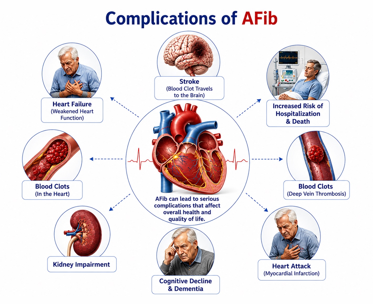
A. Thromboembolic Complications
The most feared and clinically significant complication of AFib, responsible for approximately 20–30% of all ischemic strokes and 35% of strokes in patients over 80 years. AF disrupts normal atrial blood flow dynamics, leading to stasis particularly in the left atrial appendage (LAA) — a blind-ended, trabeculated structure adjacent to the left superior pulmonary vein that is poorly contractile during AF. Stasis promotes thrombus formation according to Virchow's triad (stasis, endothelial dysfunction, hypercoagulability), and thrombus embolization to the cerebral vasculature causes large-vessel occlusion and cortical infarction.
AF-associated strokes are characteristically severe — causing large cortical infarcts with higher rates of disability, longer hospital stays, and greater mortality compared to strokes from other etiologies. The risk is not confined to sustained AF; paroxysmal AF confers equivalent per-unit-time thromboembolic risk as persistent or permanent AF. Risk stratification using CHA₂DS₂-VASc score identifies patients requiring anticoagulation regardless of AF pattern or symptom burden.
Beyond cerebral embolism, LAA thrombi may embolize to peripheral arteries — causing acute limb ischemia (femoral, popliteal, tibial occlusion), mesenteric ischemia (abdominal pain, ileus, intestinal necrosis), renal artery embolism (flank pain, hematuria, hypertension), and coronary artery embolism (acute myocardial infarction in a non-atherosclerotic vessel). These extracranial thromboembolic events occur in approximately 3.7 per 100 patient-years in non-anticoagulated AF patients, with a 5-fold reduction in risk with appropriate anticoagulation.
B. Cardiac Complications
- Heart failure and tachycardia-induced cardiomyopathy: Sustained or uncontrolled rapid ventricular rates in AF cause progressive left ventricular systolic dysfunction — a condition termed tachycardia-induced cardiomyopathy (TIC). TIC is characterized by reduced LVEF, left ventricular dilation, and symptoms of heart failure, which can be reversible with effective rate or rhythm control. Distinguishing TIC from primary cardiomyopathy is clinically important, as TIC may be fully reversible. Conversely, pre-existing heart failure promotes AF, creating a vicious cycle of mutual deterioration.
- Acute decompensated heart failure: New-onset AF or inadequately rate-controlled AF in patients with underlying diastolic or systolic dysfunction can precipitate acute pulmonary edema and hemodynamic compromise, manifesting as acute dyspnea, orthopnea, hypoxia, and pulmonary crackles. Emergency rate control or cardioversion may be required.
- Worsening of coronary artery disease: Rapid ventricular rates during AF increase myocardial oxygen demand while reducing diastolic coronary filling time, precipitating demand ischemia and potentially unstable angina or non-ST elevation myocardial infarction, particularly in patients with significant coronary stenoses.
- Sudden cardiac death: AFib modestly increases the risk of sudden cardiac death, particularly in patients with structural heart disease, Wolff-Parkinson-White syndrome (where rapid pre-excited AF can degenerate to ventricular fibrillation), or concomitant ventricular arrhythmias. The risk of ventricular fibrillation from pre-excited AF in WPW syndrome is an indication for urgent referral for accessory pathway ablation.
C. Neurological and Cognitive Complications
- Vascular dementia and cognitive decline: AF is independently associated with a 40–60% increased risk of dementia and cognitive impairment, even after controlling for overt stroke history. Mechanisms include silent cerebral microemboli producing subclinical white matter injury, chronic cerebral hypoperfusion from irregular cardiac output, and shared risk factors (hypertension, diabetes). Impaired cognitive domains include memory, executive function, processing speed, and attention.
- Depression and anxiety disorders: Affecting 20–30% of AFib patients, depression and anxiety are not merely reactive emotional responses but reflect shared neurobiological pathways involving autonomic nervous system dysregulation, inflammatory mediators, and the chronic unpredictability of paroxysmal AF episodes. Psychological comorbidity significantly worsens quality of life, reduces medication adherence, and increases healthcare utilization.
- Post-stroke depression and cognitive impairment: Survivors of AF-related strokes face disproportionately high rates of post-stroke depression, aphasia, and significant long-term functional disability, reflecting the characteristically large, cortically distributed nature of cardioembolic infarcts.
D. Complications of Antiarrhythmic and Anticoagulant Therapy
- Major bleeding from anticoagulation: The principal therapeutic risk of oral anticoagulation — including intracranial hemorrhage (most feared), gastrointestinal bleeding, and other major hemorrhage. DOAC-associated intracranial hemorrhage rates (~0.3–0.5%/year) are significantly lower than with warfarin (~0.7–1.2%/year). Bleeding risk assessment using HAS-BLED identifies modifiable risk factors (uncontrolled hypertension, concomitant antiplatelet agents, alcohol excess) that should be addressed rather than using high bleeding risk as a reason to withhold anticoagulation in high-stroke-risk patients.
- Proarrhythmia from antiarrhythmic drugs: Antiarrhythmic drugs (flecainide, propafenone, sotalol, amiodarone) carry risks of paradoxical proarrhythmic effects — including organized atrial flutter with rapid 1:1 AV conduction (flecainide/propafenone without AV nodal blocker), torsade de pointes ventricular tachycardia (sotalol, dofetilide), and ventricular proarrhythmia in structural heart disease. Class IC drugs (flecainide, propafenone) are contraindicated in structural heart disease due to the risk of ventricular proarrhythmia.
- Bradycardia and AV block: Rate-controlling agents (beta-blockers, non-dihydropyridine calcium channel blockers, digoxin) may cause excessive bradycardia, AV nodal block, and symptomatic sinus pauses — particularly when combined or in patients with underlying sinus node dysfunction or AV nodal disease.
- Amiodarone organ toxicity: Long-term amiodarone use is associated with pulmonary toxicity (pulmonary fibrosis), hepatotoxicity, thyroid dysfunction (both hyper- and hypothyroidism), corneal microdeposits, peripheral neuropathy, and photosensitivity, requiring regular organ function surveillance during therapy.
E. Quality of Life Impairment
Beyond discrete clinical endpoints, AFib profoundly impairs health-related quality of life (HRQOL) across multiple domains, affecting physical functioning, emotional well-being, and social participation. Patients with AFib report HRQOL impairment comparable to that of chronic heart failure. Persistent fatigue, exercise limitation, sleep disruption, anxiety about recurrent episodes, fear of stroke, and the burden of complex medication regimens collectively reduce functional capacity and patient well-being. Patient-reported outcome measures (PROMs) — including the AFEQT (Atrial Fibrillation Effect on Quality of Life) questionnaire and the AFSS (Atrial Fibrillation Severity Scale) — are increasingly incorporated into clinical trials and guideline-recommended care pathways.
Treatment of Atrial Fibrillation
Atrial Fibrillation (AFib) management is built upon an integrated, evidence-based four-pillar framework endorsed by ESC 2020 and AHA/ACC 2023 guidelines: (1) Anticoagulation for stroke prevention, (2) Rate control, (3) Rhythm control, and (4) Upstream therapy / Risk factor modification. Treatment must be individualized to the patient's AF type, symptom burden, hemodynamic status, comorbidities, and patient preferences.
A. Anticoagulation Therapy (Stroke Prevention)
DOACs are the preferred anticoagulants for stroke prevention in non-valvular AFib, demonstrating superior or non-inferior stroke prevention efficacy with significantly lower intracranial hemorrhage rates compared to warfarin in landmark trials:
- Apixaban (5 mg twice daily): Direct factor Xa inhibitor; demonstrated superior stroke prevention and lower bleeding compared to warfarin in ARISTOTLE trial; preferred in elderly and CKD patients due to predominantly hepatic elimination (25% renal excretion).
- Rivaroxaban (20 mg once daily with evening meal): Once-daily factor Xa inhibitor; non-inferior to warfarin in ROCKET-AF trial; convenient once-daily dosing improves adherence.
- Dabigatran (150 mg twice daily; 110 mg twice daily in elderly/high bleeding risk): Direct thrombin inhibitor; demonstrated superior stroke prevention at 150 mg dose in RE-LY trial; primarily renally cleared — contraindicated in eGFR <30 mL/min; reversible with idarucizumab.
- Edoxaban (60 mg once daily; 30 mg if <60 kg, eGFR 15–50, or P-gp inhibitors): Factor Xa inhibitor; non-inferior to warfarin with lower bleeding in ENGAGE AF trial; once-daily dosing.
All DOACs are contraindicated in valvular AF (rheumatic mitral stenosis or mechanical prosthetic heart valves) — warfarin is mandatory in these settings.
Warfarin remains the anticoagulant of choice for valvular AF (rheumatic mitral stenosis, mechanical prosthetic valves — target INR 2.5–3.5 for mechanical mitral valves). It requires regular INR monitoring (target 2.0–3.0), has numerous food and drug interactions, and wide interindividual variability requiring dose adjustment. Time in therapeutic range (TTR) >70% is associated with optimal outcomes. Reversal with vitamin K and prothrombin complex concentrate (PCC) is available for bleeding emergencies.
For patients with non-valvular AF at high stroke risk but with absolute or relative contraindications to long-term anticoagulation (prior major intracranial hemorrhage, high fall risk, severe thrombocytopenia), percutaneous transcatheter LAA occlusion using the Watchman FLX or Amulet devices provides non-pharmacological stroke prevention by mechanically sealing the LAA. LAAO is non-inferior to warfarin for stroke prevention in the PROTECT-AF and PREVAIL trials. Surgical LAA closure/ligation may be performed concomitantly during cardiac surgery.
B. Rate Control
- Target heart rate: A resting ventricular rate <110 bpm is the recommended rate control target (lenient rate control — RACE II trial), with a stricter target (<80 bpm at rest, <110 bpm with moderate exercise) for patients with persistent symptoms or heart failure with tachycardia-induced cardiomyopathy.
- Beta-blockers (first-line): Metoprolol, bisoprolol, carvedilol, and atenolol effectively reduce ventricular rate by blocking sympathetic stimulation of the AV node. Preferred in heart failure with reduced EF (carvedilol, bisoprolol, or metoprolol succinate are evidence-based for HFrEF), post-MI, and hyperthyroidism-induced AF. Side effects include fatigue, bradycardia, bronchospasm (avoid in severe asthma), and impotence.
- Non-dihydropyridine calcium channel blockers (NDHPs) — Diltiazem and Verapamil: Effective AV nodal blockers providing rate control with vasodilatory and anti-ischemic properties. Preferred in patients with obstructive lung disease (as an alternative to beta-blockers). Absolutely contraindicated in heart failure with reduced EF due to negative inotropic effects.
- Digoxin: Provides rate control through vagal enhancement and direct AV nodal effects; most effective at rest and in sedentary or elderly patients. Limited utility in physically active patients due to attenuated rate control during adrenergic activation with exercise. Narrow therapeutic index requires close serum level monitoring; toxicity (nausea, visual disturbance, arrhythmias) may occur at levels >2.0 ng/mL. Useful as add-on therapy in HFrEF patients requiring additional rate control.
- AV node ablation with pacemaker implantation ("ablate and pace"): Reserved for patients with refractory symptomatic rapid AF where pharmacological rate control fails or is not tolerated. His bundle ablation creates complete AV block, followed by permanent pacemaker implantation (preferably cardiac resynchronization therapy / CRT-D in HFrEF). Effective for symptom relief but commits the patient to permanent pacemaker dependency.
C. Rhythm Control
- Electrical cardioversion (DC cardioversion / DCCV): Synchronized direct current cardioversion — delivering a synchronized electrical shock (typically 100–200 J biphasic) during the cardiac R wave — terminates AF by simultaneously depolarizing all atrial cells, allowing the SA node to re-establish coordinated sinus rhythm. Highly effective (>90% initial success for persistent AF). Requires adequate anticoagulation: if AF duration >48 hours or unknown, therapeutic anticoagulation for ≥3 weeks before cardioversion, or TOE-guided cardioversion with exclusion of LAA thrombus, followed by at least 4 weeks of anticoagulation post-cardioversion regardless of CHA₂DS₂-VASc score (risk of stunning-related thrombus formation post-cardioversion).
- Pharmacological cardioversion: Intravenous flecainide (150–300 mg IV over 10–30 min) or propafenone are highly effective (>70%) for recent-onset AF (<48 hours duration) in patients without structural heart disease. IV amiodarone (5–7 mg/kg IV over 1–2 hours) is preferred in structural heart disease. Oral "pill-in-the-pocket" approach with flecainide (200–300 mg orally) or propafenone (450–600 mg orally) allows patient-initiated self-cardioversion of recognized paroxysmal AF episodes in appropriately selected patients after supervised testing.
- Flecainide and Propafenone (Class IC): Highly effective for maintaining sinus rhythm in patients without structural heart disease or significant coronary artery disease. Both agents block fast sodium channels, slowing atrial conduction. Contraindicated in ischemic heart disease and HFrEF due to proarrhythmic risk (CAST trial lessons). Require concurrent AV nodal blocking agent to prevent organized flutter with rapid 1:1 AV conduction.
- Sotalol (Class III, beta-blocker): Combines beta-blockade with potassium channel blockade. Moderate efficacy for AF maintenance. Risk of QT prolongation and torsade de pointes; requires baseline QTc <450 ms and eGFR >40 mL/min. Beneficial in patients with coronary artery disease and AF.
- Amiodarone (Class III — multi-channel blocker): The most effective antiarrhythmic drug for AF maintenance (>65% sinus rhythm at 1 year), safe in structural heart disease and HFrEF. However, significant organ toxicity profile (pulmonary, hepatic, thyroid, ocular, neurological) limits long-term use. Often reserved for patients with structural heart disease or where other AADs have failed, using the lowest effective dose with regular toxicity monitoring.
- Dronedarone (Class III analog of amiodarone): Less effective than amiodarone but with better tolerability profile and no thyroid/pulmonary toxicity. Contraindicated in permanent AF, NYHA Class III-IV heart failure, HFrEF, and hepatic impairment. Reduces cardiovascular hospitalization (ATHENA trial). Preferred for maintaining sinus rhythm in paroxysmal/persistent AF with minimal or no structural heart disease.
- Dofetilide (Class III): Selective IKr potassium channel blocker; safe in structural heart disease and HFrEF. Requires in-hospital initiation with continuous ECG monitoring for QT prolongation and torsade de pointes risk. Primarily used in the United States.
Catheter ablation has emerged as the most effective rhythm control strategy, particularly for symptomatic paroxysmal and persistent AF refractory to or intolerant of antiarrhythmic drug therapy:
- Pulmonary vein isolation (PVI): The cornerstone of AF ablation, based on the seminal observation (Haïssaguerre et al., 1998) that the majority of AF triggers originate from pulmonary vein myocardial sleeves. Electrical isolation of all four pulmonary veins from the left atrial body by creating circumferential ablation lesions prevents trigger-initiated AF. Techniques include radiofrequency (RF) ablation with point-by-point lesion delivery, cryoballoon ablation (Arctic Front) for en-bloc pulmonary vein isolation, pulsed-field ablation (PFA — newest energy modality with selective atrial myocyte targeting and superior safety profile for esophageal and phrenic nerve protection), and laser balloon ablation.
- PVI efficacy: Freedom from AF at 12 months post-ablation — approximately 70–80% for paroxysmal AF and 50–65% for persistent AF after a single procedure. Multiple procedures improve success rates by 10–15%. Significantly superior to antiarrhythmic drug therapy for freedom from AF recurrence in multiple randomized trials (CABANA, MANTRA-PAF, RAAFT-2).
- Extended ablation strategies: For persistent and longstanding persistent AF, adjunctive lesion sets beyond PVI — including posterior wall isolation, superior vena cava isolation, linear ablation (mitral isthmus, roof line), complex fractionated atrial electrogram (CFAE) ablation, and rotor/focal impulse ablation — are performed at experienced centers to improve outcomes in substrate-dependent AF.
- Early rhythm control (EAST-AFNET 4 trial): The landmark EAST-AFNET 4 trial demonstrated that early rhythm control initiated within 12 months of first AF diagnosis significantly reduced the composite of cardiovascular death, stroke, hospitalization for heart failure, and acute coronary syndrome compared to rate control strategy alone, establishing the principle of early rhythm control as the preferred paradigm in newly diagnosed AF.
D. Upstream Therapy and Risk Factor Modification
Addressing underlying conditions and modifiable risk factors is a cornerstone of comprehensive AFib management ("ABC" pathway — Anticoagulation/Avoid stroke, Better symptom management, and Cardiovascular risk factor/Comorbidity reduction):
- Hypertension management: Achieving blood pressure targets <130/80 mmHg with RAS-blocking agents (ACE inhibitors, ARBs) provides additional anti-fibrotic effects on atrial myocardium beyond blood pressure lowering, reducing AF recurrence. Sacubitril-valsartan (ARNI) may provide superior atrial reverse remodeling benefits.
- Weight loss: The LEGACY trial demonstrated that sustained weight loss >10% in obese patients significantly reduces AF burden, EHRA symptom class, and AF recurrence after ablation. Weight management should be actively pursued and monitored in all overweight AFib patients.
- CPAP therapy for OSA: Continuous positive airway pressure reduces AF burden in patients with comorbid OSA and improves ablation outcomes. Pre-ablation OSA screening and CPAP optimization are recommended by major guidelines.
- Alcohol cessation: Complete abstinence from alcohol in patients with AF reduces AF recurrence significantly, as demonstrated in the REDUAF randomized trial. Even reduction of alcohol intake in moderate drinkers provides measurable benefit.
- Exercise therapy: Structured aerobic exercise (cardiac rehabilitation) reduces AF symptom burden, improves quality of life, and may reduce AF recurrence. Avoidance of excessive high-intensity endurance exercise training in athletes with AF.
- Statin therapy: Beyond lipid-lowering, statins provide pleiotropic anti-inflammatory and anti-fibrotic effects that may reduce AF recurrence post-cardioversion and post-ablation, particularly in patients with structural heart disease.
- Mineralocorticoid receptor antagonists (spironolactone, eplerenone): In patients with heart failure and hypertension, MRA therapy reduces atrial fibrosis and AF burden through aldosterone receptor blockade in the atrial myocardium.
E. Emergency Management of Acute AFib
- Hemodynamically unstable AF: Immediate emergency synchronized DC cardioversion (100–200 J biphasic); concurrent IV heparin anticoagulation; treat precipitants (correct electrolytes, treat sepsis, exclude pulmonary embolism).
- Pre-excited AF (WPW syndrome): Avoid AV nodal blocking agents (digoxin, verapamil, diltiazem, adenosine) which paradoxically accelerate conduction through the accessory pathway, potentially precipitating ventricular fibrillation. Emergency cardioversion or IV procainamide/ibutilide followed by urgent accessory pathway ablation is the definitive treatment.
- Hemodynamically stable new AF (<48 hours): Rate control with IV beta-blocker or diltiazem; anticoagulation with IV/SC heparin or DOAC initiation; consider pharmacological or electrical cardioversion if symptoms persist; if duration uncertain — TOE-guided approach or 3-week anticoagulation pre-cardioversion.
- AF with acute coronary syndrome: Antithrombotic management (triple therapy — anticoagulation + dual antiplatelet therapy) is required acutely, with transition to dual therapy (anticoagulant + single antiplatelet) after 1–4 weeks to balance stroke prevention against bleeding risk.
Prevention and Control of Atrial Fibrillation
Prevention of Atrial Fibrillation (AFib) spans primary prevention (reducing incident AF by targeting modifiable risk factors in the general population), secondary prevention (preventing AF recurrence and complications in those already diagnosed), and tertiary prevention (minimizing disability and improving quality of life in established AF). The upstream therapy approach integrates all three levels.
A. Primary Prevention — Reducing Incident AFib
- Optimal blood pressure control: Hypertension is the single most prevalent modifiable AF risk factor at the population level. Achieving guideline-recommended blood pressure targets (<130/80 mmHg) through lifestyle modification and pharmacotherapy, with preference for renin-angiotensin system (RAS)-blocking agents that provide additional anti-fibrotic benefits, reduces AF incidence. Population-level hypertension control could prevent approximately 1 in 5 AF cases.
- Obesity and metabolic health management: Achieving and maintaining a healthy body weight through dietary modification and physical activity programs reduces AF risk. Structured weight management programs targeting BMI <27 kg/m² in overweight and obese individuals reduce new AF incidence. Bariatric surgery in morbidly obese patients has demonstrated significant reductions in AF incidence and recurrence.
- Moderate, regular aerobic exercise: Regular moderate-intensity aerobic exercise (150–300 minutes per week of moderate-intensity activity) is cardioprotective and reduces AF risk. Avoiding excessive high-intensity endurance exercise training (particularly >2000–3000 hours lifetime of vigorous exercise) may reduce AF risk in male athletes. Exercise prescriptions should be tailored to individual cardiovascular status.
- Alcohol reduction and cessation: Limiting alcohol consumption to <1 standard drink per day, or ideally abstaining completely in individuals at high AF risk, reduces AF incidence. Given the dose-dependent relationship between alcohol and AF, alcohol reduction is a highly impactful and underutilized preventive strategy.
- Tobacco cessation: Smoking cessation reduces AF risk over time and provides broad cardiovascular protection. Pharmacological cessation aids (varenicline, bupropion, nicotine replacement) should be offered to smoking AF patients.
- Management of sleep apnea: Screening for and treating obstructive sleep apnea with CPAP therapy reduces incident AF in high-risk populations. OSA screening (Epworth Sleepiness Scale, STOP-Bang questionnaire, polysomnography when indicated) should be incorporated into cardiovascular prevention programs.
- Diabetes and dyslipidemia management: Optimizing glycemic control (HbA1c targets), statin therapy for lipid management, and comprehensive metabolic risk factor treatment reduce the cumulative AFib risk attributable to metabolic syndrome components.
- Dietary approaches: Adherence to Mediterranean-style dietary patterns — rich in olive oil, vegetables, fruits, whole grains, fish, and nuts, with limited processed foods and added sugars — is associated with reduced AF incidence and cardiovascular risk. Adequate dietary potassium and magnesium intake supports atrial membrane stability.
B. Secondary Prevention — Preventing Recurrence and Complications
- Continued anticoagulation: Sustained anticoagulation in patients with CHA₂DS₂-VASc score ≥2 (males) / ≥3 (females) regardless of AF pattern (paroxysmal, persistent, or those who achieve apparent sinus rhythm restoration) — because stroke risk persists even during periods of apparent sinus rhythm in patients who previously had AF.
- Post-ablation risk factor management: Aggressive risk factor modification after catheter ablation significantly improves long-term freedom from AF recurrence. The ARREST-AF and LEGACY studies demonstrated that comprehensive risk factor management programs reduce post-ablation AF recurrence by 45–50% compared to standard care.
- Cardiac rehabilitation: Structured, supervised exercise and comprehensive risk factor management programs improve exercise capacity, quality of life, and cardiovascular outcomes in AFib patients, analogous to their well-established role in coronary artery disease and heart failure.
- Regular follow-up and adherence monitoring: Structured follow-up visits for medication adherence assessment, DOAC and antiarrhythmic drug dose review, symptom monitoring (EHRA scoring), blood pressure and weight targets, and detection of comorbidity progression are essential components of secondary prevention.
C. Stroke Prevention Programs
- Systematic AF screening programs: Population-based opportunistic ECG screening — particularly in individuals over 65 years — increases detection of silent AF, enabling anticoagulation initiation before the first stroke. Pulse palpation, hand-held ECG devices (KardiaMobile), and smartwatch-based PPG algorithms are scalable screening tools. Several national health systems now recommend systematic AF screening as part of preventive care programs for older adults.
- Anticoagulation adherence support: Patient education programs emphasizing the critical importance of daily anticoagulant adherence, smartphone reminder applications, blister pack dispensing, and pharmacist-led anticoagulation clinics improve adherence and reduce stroke rates in AFib populations.
- Integrated care pathways and patient empowerment: Nurse-led or pharmacist-led AFib management programs, incorporating patient education, self-monitoring, digital health tools, and structured follow-up, improve anticoagulation adherence, DOAC persistence, symptom control, and quality of life. The ESC "ABC" pathway (Avoid stroke, Better symptom management, Cardiovascular risk factor management) provides a structured template for integrated AF care.
D. Public Health and Awareness Strategies
- Community AF awareness campaigns: Public education initiatives promoting awareness of AF symptoms (palpitations, irregular pulse), risk factors, and the importance of timely medical evaluation enable earlier diagnosis and stroke prevention.
- Digital health and mHealth technologies: Smartphone-enabled ECG recording, wearable cardiac monitors, AI-powered arrhythmia detection algorithms, and telehealth platforms expand access to AF screening and monitoring beyond traditional clinical settings, particularly benefiting rural and underserved populations.
- Healthcare provider education: Training primary care physicians, pharmacists, nurses, and paramedics in AF recognition, CHA₂DS₂-VASc scoring, anticoagulation initiation, and timely referral pathways improves early detection and guideline-adherent management at the community level.
- Policy and regulatory measures: Public health policies promoting physical activity, healthy diets, alcohol reduction, tobacco control, and hypertension screening provide the population-level foundation for reducing the growing global burden of AFib.
Common FAQs on Atrial Fibrillation
Bibliography on Atrial Fibrillation
- Hindricks G, Potpara T, Dagres N, et al. 2020 ESC Guidelines for the diagnosis and management of atrial fibrillation. European Heart Journal. 2021;42(5):373–498.
- January CT, Wann LS, Calkins H, et al. 2019 AHA/ACC/HRS Focused Update of the 2014 AHA/ACC/HRS Guideline for Management of Patients With Atrial Fibrillation. Journal of the American College of Cardiology. 2019;74(1):104–132.
- Kirchhof P, Camm AJ, Goette A, et al. Early Rhythm-Control Therapy in Patients with Atrial Fibrillation. New England Journal of Medicine. 2020;383(14):1305–1316. [EAST-AFNET 4 Trial]
- Granger CB, Alexander JH, McMurray JJV, et al. Apixaban versus Warfarin in Patients with Atrial Fibrillation. New England Journal of Medicine. 2011;365(11):981–992. [ARISTOTLE Trial]
- Connolly SJ, Ezekowitz MD, Yusuf S, et al. Dabigatran versus Warfarin in Patients with Atrial Fibrillation. New England Journal of Medicine. 2009;361(12):1139–1151. [RE-LY Trial]
- Patel MR, Mahaffey KW, Garg J, et al. Rivaroxaban versus Warfarin in Nonvalvular Atrial Fibrillation. New England Journal of Medicine. 2011;365(10):883–891. [ROCKET-AF Trial]
- Calkins H, Hindricks G, Cappato R, et al. 2017 HRS/EHRA/ECAS/APHRS/SOLAECE Expert Consensus Statement on Catheter and Surgical Ablation of Atrial Fibrillation. Heart Rhythm. 2017;14(10):e275–e444.
- Pathak RK, Middeldorp ME, Lau DH, et al. Aggressive Risk Factor Reduction Study for Atrial Fibrillation and Implications for the Outcome of Ablation. Journal of the American College of Cardiology. 2014;64(21):2222–2231. [ARREST-AF Trial]
- Pathak RK, Evans M, Middeldorp ME, et al. Cost-Effectiveness and Clinical Effectiveness of the Risk Factor Management Clinic in Atrial Fibrillation. JACC: Clinical Electrophysiology. 2017;3(3):253–263. [LEGACY Study]
- Lip GYH, Nieuwlaat R, Pisters R, Lane DA, Crijns HJGM. Refining clinical risk stratification for predicting stroke and thromboembolism in atrial fibrillation using a novel risk factor-based approach. Chest. 2010;137(2):263–272. [CHA₂DS₂-VASc Score Derivation]
- Joglar JA, Chung MK, Armbruster AL, et al. 2023 ACC/AHA/ACCP/HRS Guideline for Diagnosis and Management of Atrial Fibrillation. Journal of the American College of Cardiology. 2024;83(1):109–279.
- Haïssaguerre M, Jaïs P, Shah DC, et al. Spontaneous Initiation of Atrial Fibrillation by Ectopic Beats Originating in the Pulmonary Veins. New England Journal of Medicine. 1998;339(10):659–666. [Landmark PV Triggers Paper]
- Harrison's Principles of Internal Medicine, 21st ed. Editors: Jameson JL, Fauci AS, Kasper DL, Hauser SL, Loscalzo J. McGraw-Hill Education.
- Brandes A, Smit MD, Nguyen BO, Rienstra M, Van Gelder IC. Risk factor management in atrial fibrillation. Arrhythmia & Electrophysiology Review. 2018;7(2):118–127.
- Freedman B, Potpara TS, Lip GYH. Stroke prevention in atrial fibrillation. Lancet. 2016;388(10046):806–817.