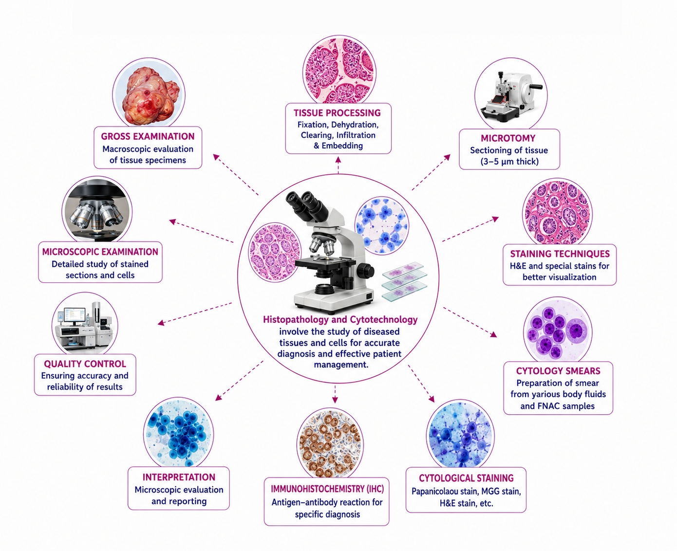
Histopathology and Cytotechnology are specialized branches of laboratory medicine concerned with the microscopic evaluation of tissues and cells for diagnostic, prognostic, and research purposes. Unlike hematology and clinical chemistry, histopathology does not primarily rely on numerical reference ranges but rather on established normal histological architecture, cellular morphology, tissue composition, staining characteristics, morphometric measurements, proliferative indices, immunohistochemical expression patterns, and cytological criteria. These reference parameters serve as the foundation for distinguishing normal physiological structures from inflammatory, infectious, degenerative, premalignant, and malignant conditions.
Histopathology Parameters
Tissue Processing Standards
Specimen Fixation Parameters
| Parameter |
Reference Value |
| Fixative |
10% Neutral Buffered Formalin |
| Formaldehyde Concentration |
4% |
| Fixative-to-Tissue Ratio |
10:1 to 20:1 |
| Minimum Fixation Time |
6 Hours |
| Optimal Fixation Time |
24–48 Hours |
| Maximum Recommended Time |
72 Hours |
| Tissue Thickness |
≤5 mm |
Tissue Processing Schedule
| Step |
Reference Range |
| Dehydration (70% Ethanol) |
1–2 Hours |
| Dehydration (95% Ethanol) |
1–3 Hours |
| Absolute Alcohol |
1–3 Hours |
| Xylene Clearing |
30–90 Minutes |
| Paraffin Infiltration |
45–120 Minutes |
| Paraffin Temperature |
56–60°C |
Histological Section Parameters
| Parameter |
Reference Value |
| Routine Section Thickness |
3–5 μm |
| Kidney Biopsy Sections |
2–3 μm |
| Bone Marrow Sections |
2–4 μm |
| Frozen Section Thickness |
5–10 μm |
| Electron Microscopy Sections |
50–100 nm |
Normal Cellular Morphometry
Nuclear Parameters
| Parameter |
Reference Value |
| Nuclear Diameter |
4–10 μm |
| Nuclear-Cytoplasmic Ratio |
1:4 to 1:6 |
| Nucleoli |
Inconspicuous |
| Chromatin Pattern |
Fine, Evenly Distributed |
| Nuclear Membrane |
Smooth and Regular |
Cellular Parameters
| Parameter |
Reference Value |
| Cell Diameter |
10–30 μm |
| Cytoplasmic Border |
Well Defined |
| Cytoplasmic Staining |
Uniform |
| Mitotic Figures |
Rare |
Normal Histological Architecture
Skin
Epidermis
| Layer |
Normal Thickness |
| Stratum Corneum |
10–20 μm |
| Epidermis (Overall) |
50–150 μm |
Dermis
| Parameter |
Reference Value |
| Thickness |
1–4 mm |
| Collagen Fibers |
Uniform Distribution |
| Elastic Fibers |
Intact |
Liver
| Parameter |
Reference Value |
| Hepatocyte Diameter |
20–30 μm |
| Hepatic Lobule Diameter |
1–2 mm |
| Binucleated Hepatocytes |
<25% |
| Portal Tracts |
Regular Distribution |
Kidney
Glomerulus
| Parameter |
Reference Value |
| Glomerular Diameter |
150–250 μm |
| Mesangial Cells |
≤3 Cells/Mesangial Area |
| Basement Membrane Thickness |
300–400 nm |
Renal Tubules
| Parameter |
Reference Value |
| Tubular Epithelium |
Intact |
| Brush Border |
Preserved |
Thyroid
| Parameter |
Reference Value |
| Follicular Diameter |
50–500 μm |
| Colloid |
Homogeneous |
| Follicular Epithelium Height |
6–10 μm |
Lung
| Parameter |
Reference Value |
| Alveolar Septal Thickness |
<10 μm |
| Alveolar Macrophages |
Occasional |
| Type II Pneumocytes |
<10% Alveolar Cells |
Gastrointestinal Tract
Small Intestine
| Parameter |
Reference Value |
| Villus Height |
300–1500 μm |
| Crypt Depth |
100–300 μm |
| Villus:Crypt Ratio |
3:1–5:1 |
Colon
| Parameter |
Reference Value |
| Crypt Length |
400–500 μm |
| Goblet Cells |
Abundant |
Bone Marrow Histopathology
| Parameter |
Reference Value |
| Cellularity (Adult) |
30–70% |
| Fat Content |
30–70% |
| Myeloid:Erythroid Ratio |
2:1–4:1 |
| Blasts |
<5% |
| Plasma Cells |
<3% |
Histochemical Staining Parameters
Hematoxylin and Eosin (H&E)
| Parameter |
Expected Appearance |
| Nuclei |
Blue–Purple |
| Cytoplasm |
Pink |
| Collagen |
Pink |
| Muscle |
Pink–Red |
Periodic Acid-Schiff (PAS)
| Structure |
Expected Staining |
| Glycogen |
Magenta |
| Basement Membrane |
Magenta |
| Fungal Cell Walls |
Positive |
Masson's Trichrome
| Structure |
Expected Staining |
| Collagen |
Blue/Green |
| Muscle |
Red |
| Nuclei |
Black |
Immunohistochemistry (IHC) Reference Expression
Proliferation Markers
| Marker |
Normal Expression |
| Ki-67 |
<5% in Most Adult Tissues |
| PCNA |
Low Basal Activity |
Epithelial Markers
| Marker |
Normal Expression |
| Pan-Cytokeratin |
Positive |
| CK7 |
Tissue Dependent |
| CK20 |
Tissue Dependent |
| EMA |
Positive |
Mesenchymal Markers
| Marker |
Normal Expression |
| Vimentin |
Positive |
| Desmin |
Muscle Cells |
| Smooth Muscle Actin |
Smooth Muscle |
Lymphoid Markers
| Marker |
Normal Expression |
| CD3 |
T Lymphocytes |
| CD20 |
B Lymphocytes |
| CD45 |
Leukocytes |
Cytotechnology Parameters
Cytotechnology involves microscopic evaluation of exfoliated cells, body fluid cytology, fine needle aspiration cytology (FNAC), and gynecological cytology.
Cervicovaginal Cytology (Bethesda System)
Normal Squamous Cells
| Parameter |
Reference Value |
| Cell Diameter |
30–60 μm |
| Nucleus Diameter |
4–8 μm |
| N:C Ratio |
Low |
| Chromatin |
Fine |
Intermediate Cells
| Parameter |
Reference Value |
| Diameter |
30–50 μm |
| Cytoplasm |
Abundant |
| Nucleus |
Central |
Superficial Cells
| Parameter |
Reference Value |
| Diameter |
40–60 μm |
| Nucleus |
Pyknotic |
Fine Needle Aspiration Cytology (FNAC)
Adequacy Criteria
| Site |
Minimum Adequate Cells |
| Thyroid |
≥6 Cell Groups |
| Breast |
≥6 Epithelial Clusters |
| Lymph Node |
Adequate Lymphoid Population |
Body Fluid Cytology
Pleural Fluid
| Parameter |
Normal Value |
| Mesothelial Cells |
Present |
| Macrophages |
Few |
| Neutrophils |
Rare |
| Malignant Cells |
Absent |
Ascitic Fluid
| Parameter |
Normal Value |
| Mesothelial Cells |
Present |
| Reactive Cells |
Minimal |
| Tumor Cells |
Absent |
Sputum Cytology
| Parameter |
Normal Finding |
| Bronchial Cells |
Occasional |
| Macrophages |
Present |
| Squamous Cells |
Few |
| Malignant Cells |
Absent |
Urine Cytology
| Parameter |
Normal Finding |
| Urothelial Cells |
Few |
| Squamous Cells |
Occasional |
| Atypical Cells |
Absent |
| Malignant Cells |
Absent |
Cytomorphometric Parameters
| Parameter |
Normal Range |
| Nuclear Area |
20–80 μm² |
| Cytoplasmic Area |
100–500 μm² |
| Nuclear Circularity |
0.8–1.0 |
| N:C Ratio |
1:4–1:6 |
Quality Assurance Parameters
Histopathology Laboratory
| Parameter |
Standard |
| Formalin pH |
7.0–7.2 |
| Paraffin Melting Point |
56–58°C |
| Water Bath Temperature |
40–45°C |
| Slide Drying Temperature |
60°C |
Cytology Laboratory
| Parameter |
Standard |
| Alcohol Fixative |
95% Ethanol |
| Fixation Time |
≥15 Minutes |
| Pap Stain Quality |
Uniform |
| Screening Sensitivity |
>95% |
References
- Rosai and Ackerman's Surgical Pathology.
- Sternberg's Diagnostic Surgical Pathology.
- Bancroft's Theory and Practice of Histological Techniques.
- Wheater's Functional Histology.
- Koss' Diagnostic Cytology and Its Histopathologic Bases.
- Comprehensive Cytopathology.
- College of American Pathologists.
- World Health Organization.
- International Academy of Cytology.
- Clinical and Laboratory Standards Institute.