


Hepatitis C – Sign & Symptoms, Transmission, Risk factors, Diagnosis, Complication, Treatment and Prevention
Hepatitis C is a chronic blood-borne infectious disease of the liver caused by the Hepatitis C virus (HCV), a positive-sense single-stranded RNA virus belonging to the genus *Hepacivirus*, family *Flaviviridae*. Unlike Hepatitis A and B, Hepatitis C was not identified as a distinct pathogen until 1989, when it was isolated as the principal cause of non-A, non-B hepatitis following blood transfusion. It is a major global health burden: the WHO estimates approximately 58 million people have chronic HCV infection globally, with approximately 1.5 million new infections annually and approximately 290,000 deaths per year from HCV-related liver disease and hepatocellular carcinoma. Despite the availability of highly effective curative treatments since 2013, only a fraction of infected individuals have been diagnosed and treated, underscoring the critical importance of expanded screening and linkage-to-care programs.
HCV is an enveloped virus with a single-stranded, positive-sense RNA genome of approximately 9.6 kilobases encoding a single large polyprotein of approximately 3,011 amino acids that is co- and post-translationally cleaved into three structural proteins (Core, E1, E2) and seven non-structural proteins (p7, NS2, NS3, NS4A, NS4B, NS5A, NS5B). The NS3/4A serine protease and the NS5B RNA-dependent RNA polymerase (RdRp) — which lacks a proofreading mechanism — are the primary targets of direct-acting antiviral (DAA) therapies. The high error rate of the NS5B RdRp generates extraordinary genetic diversity: seven major genotypes (HCV-1 to HCV-7) with at least 86 subtypes, with significant geographic variation in genotype distribution. Genotype 1 is the most prevalent worldwide, genotype 3 predominates in South Asia, and genotype 4 in sub-Saharan Africa and the Middle East. 
HCV infects hepatocytes through a multi-step process involving attachment to host receptors including CD81, claudin-1, occludin, and SR-BI. Within hepatocytes, HCV replicates in the endoplasmic reticulum, assembling new virions that are secreted into the bloodstream. The pathophysiology of HCV-related liver injury is primarily immune-mediated: HCV-specific CD4+ and CD8+ T-cell responses target infected hepatocytes, causing hepatocytic necrosis and inflammatory infiltration. However, HCV employs multiple immune evasion strategies — including NS3/4A protease cleavage of MAVS and TRIF adaptors in innate immune signaling pathways — to establish persistent infection. In approximately 75–85% of acutely infected individuals, the virus evades immune clearance and establishes chronic infection, driving progressive hepatic fibrosis, cirrhosis, and hepatocellular carcinoma over decades.
Sign and Symptoms of Hepatitis C
Hepatitis C is frequently called the "silent epidemic" because the vast majority of acutely and chronically infected individuals are asymptomatic or have non-specific symptoms for years to decades, making clinical diagnosis extremely challenging without laboratory screening. By the time symptoms of advanced liver disease appear, significant — often irreversible — hepatic damage has already occurred.
1. Acute Hepatitis C
The incubation period of acute HCV ranges from 2 to 26 weeks, with a median of 7–8 weeks. The acute phase is asymptomatic in approximately 75–80% of individuals, which substantially limits recognition, diagnosis, and early intervention. When acute HCV does produce symptoms, they are non-specific and typically mild, rarely prompting medical evaluation.
Symptomatic Acute HCV (20–25% of Cases)
- Fatigue and Malaise: The most common symptom of acute HCV, often profound and disproportionate to objective findings. Fatigue arises from systemic interferon responses and cytokine-mediated immune activation.
- Nausea, Vomiting, and Anorexia: Gastrointestinal symptoms are common and may contribute to weight loss.
- Right Upper Quadrant Discomfort: Dull aching or heaviness in the right upper quadrant reflects hepatic capsular distension from hepatic inflammation and hepatomegaly.
- Fever and Myalgia: Low-grade fever and generalized muscle aches occur in some patients during acute viremia.
- Jaundice: Icteric acute HCV occurs in approximately 20–30% of symptomatic patients, manifesting as scleral icterus, yellow skin discoloration, dark urine (bilirubinuria), and pale stools. Importantly, paradoxically, the approximately 20–25% of patients who develop jaundice during acute HCV are more likely to spontaneously clear the infection — jaundice may represent a more vigorous immune response associated with better viral control.
- Arthralgia: Joint pain, particularly of small joints, may occur during the acute phase from immune complex deposition.
Spontaneous Resolution of Acute HCV
Approximately 15–25% of acutely infected individuals spontaneously clear HCV viremia within 6 months, typically within the first 12 weeks. Spontaneous clearance is associated with: female sex, younger age at infection, symptomatic acute illness (including jaundice), certain IL28B (interferon-lambda-3) genetic polymorphisms — particularly the CC genotype — that enhance innate immune interferon responses, and robust HCV-specific CD4+ T-cell responses. After spontaneous clearance, HCV RNA becomes undetectable but anti-HCV antibodies persist lifelong; reinfection is possible as immunity is not sterilizing.
2. Chronic Hepatitis C
Chronic HCV infection is defined as persistent HCV RNA in the blood for more than 6 months. It develops in 75–85% of acutely infected individuals and is the central clinical concern in HCV management due to its potential for progressive liver injury and systemic complications over 20–40 years.
Hepatic Manifestations
- Asymptomatic Phase (First 15–20 Years): The majority of individuals with chronic HCV remain asymptomatic or experience only vague, non-specific symptoms for the first one to two decades of infection. This silent progression underlies the large proportion of undiagnosed HCV worldwide.
- Fatigue: Chronic, persistent fatigue is the most commonly reported symptom of chronic HCV, affecting up to 50–80% of patients. It is multifactorial, arising from chronic immune activation, cytokine effects on the central nervous system (particularly IL-1β and TNF-α), sleep disturbances, depression, and possibly direct neurotoxic effects of HCV or its proteins.
- Cognitive Impairment ("Brain Fog"): Difficulties with concentration, memory, and mental clarity are frequently reported by HCV patients even in the absence of cirrhosis or hepatic encephalopathy, suggesting direct HCV neurotropism or cytokine-mediated central nervous system effects.
- Right Upper Quadrant Discomfort: A dull ache or fullness in the right upper quadrant is reported by a significant minority of patients, reflecting hepatic inflammation and capsular distension.
- Jaundice and Other Features of Advanced Liver Disease: In patients who develop cirrhosis, clinical signs of hepatic decompensation emerge: jaundice, ascites, spider naevi, palmar erythema, caput medusae, hepatic encephalopathy (confusion, asterixis), variceal bleeding, coagulopathy, and muscle wasting.
Extrahepatic Manifestations of Chronic HCV
HCV is a systemic infection with diverse extrahepatic manifestations mediated by immune complex deposition, HCV-triggered B-lymphocyte proliferation, direct viral organotropism, and secondary autoimmune activation. These extrahepatic manifestations affect approximately 40–74% of HCV patients and may be the presenting complaint:
- Mixed Cryoglobulinemia: The most clinically significant and HCV-specific extrahepatic manifestation, occurring in 25–50% of chronic HCV patients. HCV-driven B-cell proliferation produces monoclonal or oligoclonal immunoglobulins (cryoglobulins — type II or III) that precipitate in the cold and form immune complexes. Clinical syndrome: purpura (palpable purpura on lower limbs — the hallmark), arthralgia, peripheral neuropathy, glomerulonephritis (membranoproliferative GN with nephrotic/nephritic syndrome), fatigue, and Raynaud's phenomenon.
- Lymphoma: Chronic HCV-driven B-lymphocyte stimulation increases the risk of B-cell non-Hodgkin lymphoma — particularly marginal zone lymphoma and diffuse large B-cell lymphoma — by 20–35-fold. HCV treatment and cure can induce complete remission of HCV-associated indolent lymphoma.
- Diabetes Mellitus: Chronic HCV is independently associated with a 1.5–2-fold increased risk of type 2 diabetes mellitus, through HCV-mediated insulin resistance, pancreatic beta-cell dysfunction, and hepatic steatosis.
- Cardiovascular Disease: HCV infection is associated with accelerated atherosclerosis, increased risk of myocardial infarction, and carotid intima-media thickness, mediated by chronic systemic inflammation, endothelial dysfunction, and metabolic effects.
- Dermatological Manifestations: Lichen planus (oral and cutaneous), porphyria cutanea tarda (blistering photosensitive rash on sun-exposed skin), necrolytic acral erythema, and prurigo nodularis.
- Sicca Syndrome (Sjögren-like): Dry eyes and dry mouth from salivary and lacrimal gland involvement, resembling primary Sjögren syndrome.
- Thyroid Disease: Thyroiditis (both hypothyroidism and hyperthyroidism) occurs with increased frequency in HCV patients, possibly due to molecular mimicry between HCV proteins and thyroid antigens.
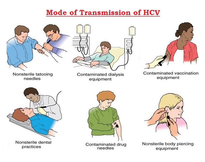
Risk Factors of Hepatitis C
A. Parenteral and Blood-Borne Risk Factors
- Injection Drug Use (IDU): Sharing of needles, syringes, cookers, cotton filters, and water among people who inject drugs (PWID) is by far the most important HCV transmission route in high-income countries, accounting for approximately 60–80% of new HCV infections. HCV RNA has been detected in dried blood on surfaces for up to 6 weeks at room temperature, enabling indirect transmission through contaminated equipment. Current injection drug use is considered the single most important modifiable risk factor for HCV.
- Blood Transfusion and Blood Products Before 1992: Before the introduction of routine HCV antibody screening of blood donors in 1992 (in most high-income countries), HCV was widely transmitted through transfusions of unscreened blood and blood products. Hemophiliacs who received pooled clotting factor concentrates before viral inactivation methods were introduced were particularly affected. In low-income countries with limited blood screening capacity, transfusion-related HCV transmission remains an ongoing risk.
- Healthcare-Associated Transmission: Unsafe injection practices (reuse of syringes and needles), inadequate sterilization of medical and dental equipment, and needlestick injuries to healthcare workers represent important HCV transmission routes in healthcare settings globally.
- Haemodialysis: Dialysis patients face elevated HCV exposure through dialysis equipment contamination, inadequate infection control, and repeated blood exposures. Prevalence of HCV among dialysis patients is several-fold higher than in the general population.
- Tattoos and Body Piercing: Procedures performed with shared, non-sterile instruments in unregulated settings carry HCV transmission risk, though the risk per procedure is lower than for injection drug use.
- Incarceration: Prisons have high HCV prevalence due to concentrated populations of PWID and opportunities for transmission through tattooing, drug use, and violence.
B. Sexual Transmission
Sexual transmission of HCV is possible but generally much less efficient than for HBV or HIV, occurring predominantly in specific high-risk contexts:
- MSM with HIV co-infection: HCV sexual transmission is significantly more common among HIV-positive MSM engaging in high-risk sexual practices (unprotected anal intercourse, fisting, use of sex toys) compared to HIV-negative MSM or heterosexual couples. Outbreaks of sexually transmitted HCV among HIV-positive MSM have been documented globally.
- Multiple sexual partners: Heterosexual transmission risk is low (approximately 0–0.6% per year in monogamous couples) but increases with multiple partners, co-existing STIs (particularly those causing mucosal ulceration), and specific sexual practices.
C. Vertical (Mother-to-Child) Transmission
Mother-to-child HCV transmission occurs in approximately 3–5% of infants born to HCV RNA-positive mothers, rising to 10–20% in mothers with HIV co-infection (due to higher HCV viral loads and compromised immune function). Unlike HBV, no effective perinatal prophylaxis is currently available for HCV. Caesarean section has not been consistently shown to reduce transmission risk. Breastfeeding is generally considered safe unless the mother has cracked or bleeding nipples.
D. Host and Genetic Risk Factors
- IL28B Genotype: The IL28B (interferon-lambda-3/IFNL3) gene polymorphism significantly influences both spontaneous HCV clearance and response to interferon-based treatment. Individuals with the CC genotype (at rs12979860) have approximately 3-fold higher rates of spontaneous clearance and superior interferon treatment response compared to TT genotype individuals.
- HIV Co-infection: HIV co-infection accelerates HCV-related liver fibrosis progression 2–3-fold and increases the risk of cirrhosis, HCC, and liver-related mortality, through impaired HCV-specific immunity, immune activation, and antiretroviral hepatotoxicity.
- Alcohol Use Disorder: Heavy alcohol consumption (>50 g/day) dramatically accelerates HCV-related fibrosis progression, increases the risk of cirrhosis 2–3-fold, and reduces the efficacy of antiviral therapy.
- Male Sex: Men with chronic HCV have faster fibrosis progression and higher HCC risk than women, possibly due to differences in sex hormone effects on hepatic stellate cell activation.
- Age at Infection and Infection Duration: Older age at the time of HCV acquisition and longer duration of infection are associated with faster fibrosis progression and higher risk of advanced liver disease.
Diagnosis of Hepatitis C
Given the largely asymptomatic nature of chronic HCV infection, population-based screening is the cornerstone of HCV diagnosis. The CDC, WHO, and most national guidelines recommend universal HCV screening for adults 18–79 years (in the United States) or for high-risk groups, combined with targeted screening of specific risk populations (PWID, recipients of blood transfusions before 1992, HIV-positive individuals, incarcerated persons, birth cohort 1945–1965 in the US).
1. Clinical Examination
- Risk Assessment and History
A comprehensive risk factor assessment should be performed in all patients, including history of injection drug use (past or current), blood transfusion or transplantation before 1992, haemodialysis, incarceration, tattoos or body piercing in unregulated settings, sexual history (MSM, multiple partners), HIV status, and birth cohort (1945–1965 in the US, where HCV prevalence peaks). A significant proportion of HCV-positive individuals have no identifiable risk factor, reinforcing the need for universal screening.
- Physical Examination
Physical examination in chronic HCV may be entirely normal in the absence of advanced liver disease. In established cirrhosis, findings include: hepatomegaly (often with left lobe predominance in cirrhosis), splenomegaly (portal hypertension), ascites, jaundice, spider naevi (>5 above the nipple line suggests cirrhosis), palmar erythema, leukonychia (white nails from hypoalbuminemia), Dupuytren's contracture, gynaecomastia, testicular atrophy, peripheral oedema, and signs of hepatic encephalopathy (asterixis, confusion, altered consciousness). Examination of the skin for purpura (lower limb), lichen planus lesions, and signs of porphyria cutanea tarda (blistering on dorsum of hands) may reveal extrahepatic manifestations.
2. Serological Diagnosis
- Anti-HCV Antibody (Screening Test)
The anti-HCV antibody test (ELISA, chemiluminescent immunoassay) is the recommended first-line screening test for HCV infection. Anti-HCV antibodies typically become detectable 8–12 weeks after infection (though third-generation assays detect antibodies as early as 4–6 weeks) and persist indefinitely, even after spontaneous viral clearance or successful treatment. A positive anti-HCV result indicates current or past HCV infection and requires confirmatory HCV RNA testing to distinguish:
- Active infection (HCV RNA positive): Current chronic or acute HCV infection requiring treatment
- Resolved infection (HCV RNA negative): Spontaneously cleared or previously treated and cured HCV
In immunocompromised patients (HIV infection, haemodialysis, organ transplantation), anti-HCV may be falsely negative despite active HCV infection; direct HCV RNA testing is recommended in these populations.
- Rapid Anti-HCV Tests
Point-of-care rapid HCV antibody tests (OraQuick HCV Rapid Antibody Test, INSTI HCV Antibody Test) on fingerstick blood or oral fluid provide results in 20–40 minutes with sensitivity and specificity comparable to laboratory-based ELISAs. They are valuable for expanding HCV testing in community settings, harm reduction programs, prisons, and resource-limited environments where laboratory infrastructure is unavailable.
3. Molecular Diagnosis
- HCV RNA Testing (Confirmatory and Monitoring)
A. Quantitative HCV RNA PCR : (viral load) is the essential confirmatory test for active HCV infection in anti-HCV-positive individuals and the primary monitoring tool during and after antiviral treatment:
- Diagnosis: Detects HCV RNA from approximately 1–2 weeks after infection (before antibodies develop), enabling diagnosis during the acute phase and in seronegative immunocompromised patients.
- Pre-treatment baseline: Quantifies viral load (in IU/mL) to guide treatment duration in some regimens.
- Treatment monitoring: HCV RNA is measured at baseline, week 4 (rapid virological response — RVR), end of treatment (ETR), and 12 weeks after treatment completion (sustained virological response — SVR12, defining cure).
- SVR12: Undetectable HCV RNA 12 weeks after completing treatment defines virological cure. SVR12 is durable in >99% of patients and is associated with normalization of liver inflammation, fibrosis regression, dramatically reduced risk of cirrhosis and HCC, and reduction in extrahepatic manifestations.
B. HCV Genotyping : HCV genotype (1–7) and subtype (e.g., 1a, 1b, 3a) determination was essential for selecting appropriate DAA regimens and predicting treatment duration with older interferon-based and first-generation DAA regimens. Pangenotypic DAA regimens (Sofosbuvir/Velpatasvir, Glecaprevir/Pibrentasvir) now eliminate the need for genotyping in most clinical scenarios, though genotyping remains useful in complex cases with prior treatment failure or suspected resistance.
4. Assessment of Hepatic Fibrosis
Fibrosis staging guides treatment urgency (patients with advanced fibrosis require priority treatment), follow-up intensity (cirrhotic patients require HCC surveillance), and identification of patients at risk for portal hypertension.
- Liver Biopsy: Histological gold standard using METAVIR scoring (F0: no fibrosis, F1: portal fibrosis, F2: periportal fibrosis, F3: bridging fibrosis, F4: cirrhosis). Increasingly replaced by non-invasive methods.
- Transient Elastography (FibroScan): Liver stiffness measurement in kPa; excellent diagnostic accuracy for significant fibrosis (F≥2, cut-off ~7 kPa) and cirrhosis (F4, cut-off ~12.5 kPa for HCV).
- FIB-4 Index: Calculated as [Age × AST] ÷ [Platelet count × √ALT]; FIB-4 <1.30 excludes significant fibrosis; FIB-4 >2.67 suggests advanced fibrosis/cirrhosis. Simple, widely available, and recommended as the first-line non-invasive fibrosis assessment by AASLD/IDSA guidelines.
- APRI Score: AST-to-Platelet Ratio Index; validated for fibrosis assessment, particularly in resource-limited settings recommended by WHO.
Complications of Hepatitis C
A. Liver Cirrhosis
Approximately 15–30% of individuals with chronic HCV develop cirrhosis over 20–30 years. Fibrosis progression is highly variable and influenced by alcohol use, age at infection, male sex, obesity, diabetes, and HIV co-infection. Cirrhosis represents the most important determinant of HCV-related mortality. Decompensated cirrhosis is defined by the occurrence of ascites, variceal bleeding, hepatic encephalopathy, or hepatorenal syndrome, carrying a 5-year mortality exceeding 80% without liver transplantation.
- Ascites : Ascites — accumulation of fluid in the peritoneal cavity — is the most common complication of HCV cirrhosis, resulting from portal hypertension and sodium and water retention from activation of the renin-angiotensin-aldosterone system. Management includes sodium restriction (<2 g/day), diuretics (spironolactone 100–400 mg/day ± furosemide 40–160 mg/day), and large-volume paracentesis with albumin infusion for refractory ascites.
- Variceal Haemorrhage : Portal hypertension drives the formation of porto-systemic venous collaterals — particularly oesophageal and gastric varices — which may rupture, causing massive upper gastrointestinal bleeding with haematemesis and melaena. Acute variceal haemorrhage is a life-threatening emergency with 6-week mortality of 15–25%, requiring urgent endoscopic variceal ligation (EVL), vasoactive drugs (terlipressin, octreotide), and antibiotic prophylaxis.
- Hepatic Encephalopathy : Hepatic encephalopathy — a neuropsychiatric syndrome ranging from subtle cognitive impairment (covert HE) to overt confusion, stupor, and coma — results from impaired hepatic clearance of ammonia and other neurotoxins. Precipitants include gastrointestinal bleeding, infection, constipation, renal failure, and electrolyte disturbances. Management includes lactulose, rifaximin, dietary protein optimization, and correction of precipitating factors.
B. Hepatocellular Carcinoma (HCC)
HCV cirrhosis carries an annual HCC incidence of 3–6%, making it a major risk factor for liver cancer. Unlike HBV, HCV does not integrate into the host genome; HCC in HCV occurs almost exclusively in the setting of cirrhosis. HCV-driven mechanisms of oncogenesis include NS5A-mediated Wnt/β-catenin and PI3K/AKT pathway activation, HCV core protein-mediated oxidative stress, and chronic inflammation-driven mutagenesis. DAA therapy significantly reduces but does not eliminate HCC risk — surveillance with 6-monthly liver ultrasound ± AFP must continue lifelong in cirrhotic patients even after achieving SVR.
C. Cryoglobulinaemic Vasculitis
Mixed cryoglobulinaemia from HCV can progress to systemic vasculitis with purpura, glomerulonephritis (causing renal failure), peripheral neuropathy (sensorimotor), and systemic involvement. DAA-induced HCV cure resolves cryoglobulinaemia in the majority of patients, with resolution of clinical manifestations over months to years.
D. HCV-Associated B-Cell Lymphoma
Chronic HCV stimulates B-lymphocyte proliferation through E2 protein interaction with CD81 on B cells, predisposing to clonal expansion and malignant transformation. HCV eradication can induce complete remission of indolent HCV-associated lymphoma (particularly splenic marginal zone lymphoma) without chemotherapy, underscoring the causal relationship.
Treatment of Hepatitis C
The treatment landscape for hepatitis C has been transformed by the introduction of Direct-Acting Antivirals (DAAs) since 2013 — oral, well-tolerated regimens achieving cure (SVR12) rates exceeding 95–99% in all HCV genotypes and disease stages, including cirrhosis and HIV co-infection, with treatment durations of only 8–12 weeks. This represents one of the greatest therapeutic revolutions in modern medicine, transforming HCV from a difficult-to-treat, incurable chronic infection to a curable disease within weeks.
A. Direct-Acting Antiviral (DAA) Therapy — First-Line
DAAs target specific steps in the HCV replication cycle through three main drug classes:
- NS5A Inhibitors : NS5A inhibitors (ending in "-asvir") block the NS5A protein, which is essential for HCV RNA replication complex formation and virion assembly. They are highly potent with picomolar EC50 values against most genotypes but have a low genetic barrier to resistance individually.
- Velpatasvir, Ledipasvir, Pibrentasvir, Elbasvir, Ombitasvir, Daclatasvir
- NS5B Polymerase Inhibitors :
- Nucleoside/Nucleotide Analogue NS5B Inhibitors (NIs): Sofosbuvir (SOF) is the prototypical NI — a uridine nucleotide prodrug that terminates HCV RNA chain elongation. It has pangenotypic activity and a high genetic barrier to resistance, serving as the backbone of most modern DAA regimens.
- Non-Nucleoside NS5B Inhibitors (NNIs): Dasabuvir targets the thumb domain of NS5B; genotype-specific and with lower genetic barrier to resistance.
- NS3/4A Protease Inhibitors : NS3/4A protease inhibitors (ending in "-previr") block the HCV serine protease, preventing polyprotein processing. They have variable genotypic coverage and genetic barrier to resistance.
- Glecaprevir, Grazoprevir, Voxilaprevir, Paritaprevir (boosted with ritonavir)
B. Recommended Pangenotypic DAA Regimens (WHO/AASLD/EASL Guidelines)
- Sofosbuvir/Velpatasvir (SOF/VEL — Epclusa) : Pangenotypic regimen (all genotypes 1–6); 400/100 mg once daily for 12 weeks. SVR12 rates >95% across all genotypes. For genotype 3 with cirrhosis, SOF/VEL/Voxilaprevir (Vosevi, 12 weeks) or SOF/VEL + ribavirin (12 weeks) is recommended. Well tolerated; minimal drug interactions.
- Glecaprevir/Pibrentasvir (GLE/PIB — Mavyret) : Pangenotypic regimen; 3 tablets once daily for 8 weeks (treatment-naive, without cirrhosis) or 12 weeks (with compensated cirrhosis). SVR12 rates >97–99% across all genotypes. Cannot be used in decompensated cirrhosis (Child-Pugh B/C) due to NS3 protease inhibitor hepatotoxicity risk. The 8-week pangenotypic regimen makes it one of the shortest-course treatment options available.
- Sofosbuvir/Ledipasvir (SOF/LDV — Harvoni) : Primarily used for HCV genotypes 1, 4, 5, and 6; 400/90 mg once daily for 8–12 weeks. SVR12 rates >95% in genotype 1. Not effective for genotype 2 or 3.
- Sofosbuvir + Daclatasvir : An alternative generic pangenotypic regimen of particular importance in low- and middle-income countries due to the widespread availability of generic formulations at dramatically lower cost. SOF 400 mg + DCV 60 mg once daily for 12 weeks (24 weeks for genotype 3 with cirrhosis). SVR12 rates >90% across genotypes.
C. Special Populations
- Cirrhosis: Compensated cirrhosis (Child-Pugh A) can be treated with standard DAA regimens with 12-week duration. Decompensated cirrhosis (Child-Pugh B/C) requires NS5B inhibitor-based regimens (SOF/VEL ± ribavirin) avoiding NS3 protease inhibitors; liver transplantation evaluation is indicated.
- HIV/HCV Co-infection: DAA regimens are highly effective in HIV/HCV co-infected patients. Careful assessment of drug-drug interactions between DAAs and antiretrovirals (particularly with cobicistat- or ritonavir-boosted regimens) is essential.
- Chronic Kidney Disease (CKD) / Renal Failure: Sofosbuvir (renally cleared metabolite accumulates) requires dose adjustment or avoidance in severe CKD (eGFR <30 mL/min); glecaprevir/pibrentasvir (primarily biliary excretion) is the preferred pangenotypic regimen for HCV patients with severe CKD or haemodialysis.
- Pregnancy: No DAA has yet received approval for use in pregnancy; women of childbearing potential should be counselled and DAA therapy deferred until after delivery if possible.
D. Post-Treatment Follow-Up
After achieving SVR12 (cure), follow-up includes:
- Cirrhotic patients: Lifelong 6-monthly HCC surveillance (ultrasound ± AFP), upper endoscopy for variceal surveillance, and liver function assessment; portal hypertension may regress slowly after SVR.
- Non-cirrhotic patients: Annual ALT and non-invasive fibrosis assessment to confirm disease stability.
- All cured patients should be counselled about risk of HCV reinfection if high-risk behaviors continue.
Prevention of Hepatitis C
Unlike Hepatitis A and B, there is no vaccine available for Hepatitis C, making prevention entirely reliant on behavioral modifications, harm reduction, infection control, and blood safety measures.
A. Harm Reduction for People Who Inject Drugs (PWID)
- Needle and Syringe Programs (NSPs): Provision of sterile injecting equipment (needles, syringes, cookers, cotton filters, water) to PWID dramatically reduces HCV (and HIV) transmission. NSPs are cost-effective, do not increase drug use, and have been associated with >70% reductions in HCV incidence in implementation studies.
- Opioid Substitution Therapy (OST): Methadone maintenance therapy and buprenorphine therapy reduce injection frequency and needle sharing, independently reducing HCV transmission risk by 50–60%.
- Drug Consumption Rooms: Supervised drug consumption facilities provide sterile equipment and prevent HCV transmission associated with drug use in unsupervised settings.
B. Blood Safety
- Universal screening of blood, organ, tissue, and cell donors for anti-HCV antibody and HCV RNA (nucleic acid testing — NAT) has effectively eliminated transfusion-transmitted HCV in countries with comprehensive screening programs.
- Mandatory use of sterile single-use syringes and needles in healthcare settings.
- Proper sterilization and disinfection of medical and dental equipment.
C. Healthcare Worker Protection
- Strict adherence to standard (universal) precautions — gloves, safe sharp disposal, avoidance of recapping needles.
- Immediate management of needlestick injuries: wound cleaning, risk assessment, testing of source patient (with consent), post-exposure monitoring of healthcare worker (HCV RNA at 6 weeks, 3 months, 6 months).
- No effective post-exposure prophylaxis is currently available for HCV; prompt identification of infection and early treatment with DAAs if seroconversion occurs.
D. Prevention of Sexual Transmission
- Condom use for individuals with multiple sexual partners or engaging in high-risk sexual practices.
- Regular HCV testing for HIV-positive MSM and others at ongoing risk.
- Partner testing and notification for sexual contacts of HCV-positive individuals.
- Treatment as prevention: curing HCV in infected individuals eliminates their infectious potential, contributing to population-level transmission reduction.
E. Treatment as Prevention
The availability of curative DAA therapy creates a unique opportunity for micro-elimination and population-level prevention. Treating HCV-infected individuals — particularly among high-risk populations such as PWID — reduces the infectious pool and HCV incidence in the community. Models project that with sufficient treatment scale-up, HCV elimination (as defined by WHO's 2030 targets of 90% reduction in incidence and 65% reduction in mortality) is achievable.
Common FAQs on Hepatitis C
- What causes Hepatitis C?
Hepatitis C is caused by the Hepatitis C virus (HCV), a positive-sense single-stranded RNA virus of the family Flaviviridae. Seven major genotypes (HCV-1 to HCV-7) with at least 86 subtypes exist globally, with genotype 1 being the most prevalent worldwide. HCV infects hepatocytes and establishes persistent infection in 75–85% of exposed individuals, driving chronic liver inflammation and progressive fibrosis.
- How is Hepatitis C transmitted?
HCV is transmitted primarily through blood-to-blood contact. The most common routes are: sharing needles, syringes, or other drug injection equipment; receiving blood transfusions or blood products before 1992 (before routine HCV screening); needlestick injuries in healthcare workers; haemodialysis; and tattoos or piercing with non-sterile equipment. Sexual transmission is possible but much less efficient than for HBV or HIV, occurring predominantly in HIV-positive MSM or individuals with multiple high-risk sexual partners.
- Can Hepatitis C be cured?
Yes — Hepatitis C is now highly curable with modern Direct-Acting Antiviral (DAA) therapy, which achieves sustained virological response (SVR12, equating to cure) in over 95–99% of patients across all genotypes with only 8–12 weeks of once-daily oral tablets. This represents a revolutionary advance compared to the 40–80% cure rates and 48-week treatment duration with older interferon-based regimens. After achieving SVR12, HCV RNA remains undetectable and does not return in >99% of patients.
- What is SVR and what does it mean?
SVR (Sustained Virological Response) — specifically SVR12 — is defined as undetectable HCV RNA in the blood 12 weeks after completing antiviral treatment. SVR12 equates to virological cure: the virus has been eradicated from the body. After SVR12, liver inflammation resolves, fibrosis may partially regress (even in cirrhosis), HCC risk significantly decreases, and extrahepatic manifestations improve. Reinfection is possible if high-risk behaviors continue.
- Why is Hepatitis C called the "silent epidemic"?
Hepatitis C is known as the "silent epidemic" because approximately 75–80% of acute HCV infections are asymptomatic, and chronic HCV causes no or only vague non-specific symptoms for one to two decades while progressive liver damage occurs silently. By the time symptoms of cirrhosis or liver failure appear, significant — often irreversible — liver damage has occurred. This explains why millions of HCV-infected individuals remain undiagnosed worldwide.
- Is there a vaccine for Hepatitis C?
No. Unlike Hepatitis A and B, there is currently no vaccine available for Hepatitis C. The extraordinary genetic variability of HCV — seven genotypes with multiple subtypes and extremely rapid mutation — has made vaccine development very challenging, as any vaccine must induce protective immunity against highly diverse viral strains. Prevention relies entirely on harm reduction, blood safety, infection control, and expanding treatment access.
- What are the symptoms of chronic Hepatitis C?
Most individuals with chronic HCV are asymptomatic or have non-specific symptoms for decades. The most common reported symptoms are chronic fatigue (affecting up to 80% of patients), mild right upper quadrant discomfort, cognitive difficulties ("brain fog"), and depression. Symptoms of advanced liver disease — jaundice, ascites, bleeding from varices, and confusion from hepatic encephalopathy — only appear when cirrhosis develops, which may take 20–30 years.
- Who should be tested for Hepatitis C?
Testing is recommended for: all adults aged 18–79 years (universal screening in the US); individuals who have ever injected drugs; people born between 1945 and 1965 (US birth cohort with high HCV prevalence); recipients of blood transfusions or organ transplants before 1992; HIV-positive individuals; people on haemodialysis; persons with unexplained elevated liver enzymes; healthcare workers after needlestick exposure; and sexual partners of HCV-positive individuals.
- How does Hepatitis C cause liver cancer?
HCV causes hepatocellular carcinoma (HCC) through chronic inflammation-mediated oxidative stress and hepatocyte damage, sustained hepatocyte regeneration (increasing mutagenesis risk), direct oncogenic effects of HCV proteins (NS5A, Core) on cell cycle regulation and signaling pathways, and mitochondrial damage. Unlike HBV, HCV-related HCC occurs almost exclusively in the setting of cirrhosis, with an annual HCC incidence of 3–6% in HCV cirrhosis.
- Can Hepatitis C be transmitted through food, water, or casual contact?
No. HCV is not transmitted through food, water, air, casual contact (hugging, shaking hands), shared utensils or glasses, coughing, sneezing, or breastfeeding (in the absence of cracked nipples). HCV requires direct blood-to-blood contact for transmission. Sharing personal items that may have blood on them (razors, toothbrushes, nail scissors) carries a theoretical transmission risk and should be avoided.
- What is the difference between Hepatitis B and Hepatitis C?
Key differences include: causative agent (HBV is a DNA virus; HCV is an RNA virus); transmission (HBV more easily sexually transmitted and perinatal; HCV primarily blood-borne); chronicity (HBV chronic in >90% of neonates, ~5% of adults; HCV chronic in ~75–85% regardless of age); vaccine availability (highly effective HBV vaccine exists; no HCV vaccine); and curability (HBV not curable but controllable; HCV now curable with DAAs in >95% of patients).
- What extrahepatic conditions are associated with Hepatitis C?
HCV is associated with numerous extrahepatic manifestations including: mixed cryoglobulinaemia (causing purpura, arthralgia, glomerulonephritis, neuropathy), B-cell non-Hodgkin lymphoma, type 2 diabetes mellitus, cardiovascular disease, lichen planus, porphyria cutanea tarda, Sjögren-like syndrome, thyroid disorders (both hypo- and hyperthyroidism), and cognitive impairment.
- How does DAA therapy work?
Direct-Acting Antivirals (DAAs) target specific HCV non-structural proteins essential for viral replication: NS5A inhibitors (e.g., velpatasvir, pibrentasvir) block the replication complex and virion assembly; NS5B nucleotide inhibitors (sofosbuvir) terminate viral RNA chain elongation; and NS3/4A protease inhibitors (e.g., glecaprevir, voxilaprevir) prevent polyprotein processing. Modern pangenotypic DAA combinations achieve >95–99% cure rates across all HCV genotypes with 8–12 weeks of once-daily oral therapy.
- Does curing Hepatitis C reverse liver damage?
Achieving SVR (cure) leads to resolution of hepatic inflammation and can produce significant fibrosis regression, including in patients with established cirrhosis. However, the degree of regression depends on the severity of pre-treatment fibrosis: patients with early fibrosis (F1–F2) typically experience near-complete normalization; those with cirrhosis may have partial fibrosis regression but portal hypertension and HCC risk (though substantially reduced) persist, requiring ongoing surveillance.
- Can a person get Hepatitis C more than once?
Yes. Successful treatment with DAAs or spontaneous clearance of HCV does not confer lasting protective immunity, unlike Hepatitis A and B. Individuals can be reinfected with the same or different HCV genotype if they are re-exposed through risk behaviors. This is particularly relevant for PWID and HIV-positive MSM with ongoing risk. Cured individuals should be retested annually or after potential re-exposure.
Bibliography
- Harrison's Principles of Internal Medicine — Editors: J. L. Jameson, A. S. Fauci, D. L. Kasper, S. L. Hauser, and J. Loscalzo. Publisher: McGraw-Hill Education.
- Mandell, Douglas, and Bennett's Principles and Practice of Infectious Diseases — Editors: J. E. Bennett, R. Dolin, and M. J. Blaser. Publisher: Elsevier.
- Sleisenger and Fordtran's Gastrointestinal and Liver Disease — Editors: M. Feldman, L. S. Friedman, and L. J. Brandt. Publisher: Elsevier.
- Oxford Textbook of Medicine — Editors: J. Firth, C. Conlon, and T. Cox. Publisher: Oxford University Press.
- AASLD-IDSA HCV Guidance: Recommendations for Testing, Managing, and Treating Hepatitis C — www.hcvguidelines.org.
- EASL Recommendations on Treatment of Hepatitis C: Final Update of the Series. Journal of Hepatology, 2020.
- WHO Guidelines on Hepatitis B and C Testing — World Health Organization, Geneva, 2017.
- Pawlotsky J.-M. et al. EASL Recommendations on Treatment of Hepatitis C. Journal of Hepatology, 2018.
- Ghany M. G. et al. Hepatitis C Guidance 2019 Update. Hepatology, 2020.
- Collaborators GH. Global Prevalence and Genotype Distribution of Hepatitis C Virus Infection in 2015. Journal of Hepatology, 2017.
- Younossi Z. et al. Global Epidemiology of Hepatitis C Virus Infection. Clinical Gastroenterology and Hepatology, 2019.
- Feld J. J. and Hoofnagle J. H. Mechanism of Action of Interferon and Ribavirin in Treatment of Hepatitis C. Nature, 2005.
- Medical Microbiology — Authors: P. R. Murray, K. S. Rosenthal, and M. A. Pfaller. Publisher: Elsevier.
- Sherris Medical Microbiology — Authors: K. J. Ryan and C. G. Ray. Publisher: McGraw-Hill Education.
- Razavi H. et al. Chronic Hepatitis C Virus (HCV) Disease Burden and Cost in the United States. Hepatology, 2013.